1Dr. KNS Memorial Institute of Medical Sciences Barabanki, Uttar Pradesh, India, 2Private Practitioner, Vitraag Dental Clinic, Shahibaug, Ahmedabad, Gujarat, India
Address for correspondence: Dr. Isha Rastogi, Dr. KNS Memorial Institute of Medical Sciences Barabanki, Uttar Pradesh – 225 001, India. E-mail: excellent123@gmail.com
Submitted: 11-Jan-2023
Accepted in Revised Form: 16-Feb-2024
Published: 03-Apr-2024
DOI: 10.4103/sidj.sidj_3_23
Saint Int Dent J 2023;7:51-4.
Copyright: © 2024 The Saint's International Dental Journal
Licence
This is an open access journal, and articles are distributed under the terms of the Creative Commons Attribution-NonCommercial-ShareAlike 4.0 License, which allows others to remix, tweak, and build upon the work non-commercially, as long as appropriate credit is given and the new creations are licensed under the identical terms.
Disclaimer:
This article was originally published by Wolters Kluwer Medknow Publications & Media Pvt Ltd and has now been officially transferred to Society of Dental Research & Education due to a change in publishing entity.
Introduction: Managing and treating severely resorbed ridges is a challenge for the dentist. Especially, in older geriatric patients who have this problem. Till now, the classification by Neil is well accepted by all for the distolingual vestibule region and lingual area.
Materials and Methods: We studied 500 patients with the presence of mandibular second molar teeth aged 20–50 years. The mirror method and a special instrument were used to view the section of the distolingual aspect present in the lingual area as well as the oral cavity.
Results: The records were seen and studied well for the class and lingual region present.
Conclusion: The distolingual alveolar sulcus and lingual region in the mandible are a guide for the tray to be used. We recorded this region in the patients by our technique.
Keywords: Distolingual alveolar sulcus, lingual, mandible
As people age, they often face both dental and medical challenges. One such challenge is the resorbed mandible and the tongue, which can make it difficult to create successful dentures for patients with high expectations.[1,2] While dental implants may be an option, financial burden and the smaller mandible denture bearing area, along with preexisting medical conditions, can be a barrier.[3,4] To prevent over-trimming of the lower denture’s lingual borders,[5,6] the lateral throat form must be properly measured during the preimpression stage. This allows for a favorable inclined plane that creates vectors of force, helping to keep the lower denture intact and improve retention and stability.[7,8] The lower denture’s distolingual flange must be properly long and wide in the distolingual sulcus to achieve a perfect peripheral seal in the lower complete denture. Muscle attachments in this area include the mylohyoid ridge anteriorly, a pear-shaped pad laterally, the superior constrictor muscle posterolaterally, the palatoglossus posteromedially, and the tongue medially.[9,10]
A total of 500 dentate subjects between 20 and 50 years were selected from the local population who came to a dental hospital. They had permanent second molars, proper mouth opening, with good neuromuscular coordination. Those with congenital abnormality or anomaly were excluded. Neil’s method of classification by mirror was used to determine the classification inside the mouth and the lingual region was seen. (Class I was no movement in the mouth mirror was seen when the tongue protruded, class II was half as long and narrow as class I flange and twice as class III, and class III was entire mirror displaced with flange 2-3mm below at mylohyoid ridge .) Then, a special instrument that had measurements was used to record the lateral throat form inside the patient’s oral cavity. Instruments used were mandibular rim lock perforated dentulous trays (SSWhite Instrument Impression Trays, Germany), a vibrator (Sirio Dental SNC, Model 47014, Italy), and a designed instrument to verify the class of lateral throat form.
It is crucial to determine the depth and classification of the lateral throat form to choose the right stock tray for an accurate preliminary impression. This leads to a good preliminary cast, followed by a well-fitted custom tray, and ultimately a stable and retentive mandibular denture. The shape of the distolingual flange helps guide the tongue on top of the denture’s flange without impeding soft tissue function. This enhances the retention and stability of the mandibular complete denture.[11]
Tongue position classification by Wright—it is class I— tongue fills the floor of the mouth. The floor of the tongue is at a higher level to lower the lingual flange of the denture producing a favorable border seal, class II—tongue is retracted and very tense, pulled backward and upward, and class III—tongue is low about mandibular ridge crest or retarded. Classes II and III are unfavorable with poor retention because they give inadequate lingual seal, overextended flanges, and dislodge dentures in tongue movements. We see that the tongue influences strongly the mandibular denture prosthesis. it is examined clinically by asking the patient to open their mouth just wide for food in small portions and inspect the different tongue positions. Martonet emphasized that lower denture stability depends upon tongue position as it is a more powerful muscle than cheeks and lips, it exerts force of 16.4 Psi.[11-15]
Discrete (categorical) data were summarized in number (n) and percentage (%) and compared by chi-square (χ2) test. A two-tailed (α = 2). P < 0.05 was considered statistically significant. Analysis was performed on SPSS software (windows version 22.0, IBM SPSS Inc., Chicago, IL, USA).
A total of 500 subjects were recruited to study the distolingual vestibule and lingual aspects (i.e., intraoral tongue position). Of these, 210 (42.0%) subjects were 20–35 years aged, and 290 (58.0%) subjects were 36–50 years aged [Table 1]. The frequency (%) of higher aged subjects (36–50 years) were 16.0% higher than lower aged subjects (20–35 years). Furthermore, among subjects, 245 (49.0%) were males, and 255 (51.0%) were females. The frequency of females was 2.0% higher than males. According to age, there were 95 (45.2%) males and 115 (54.8%) females in the 20–35 years age group, and 150 (51.7%) males and 140 (48.3%) females in the age group of 36–50 years [Table 2]. The frequency distribution of age and gender were statistically similar (χ2 = 2.05, P= 0.152). In conclusion, the study participants had higher age and female predominance.
Table 1. Distribution of age of recruited subjects
| Age (years) | Total subjects (n = 500) (%) |
|---|---|
| 20-35 | 210 (42.0) |
| 36-50 | 290 (58.0) |
Table 2. Age and gender distribution of recruited subjects
| Gender | Age | χ2 value | P value | |
|---|---|---|---|---|
| 20-35 years (n = 210) (%) | 36-50 years (n = 290) (%) | |||
| Male | 95 (45.2) | 150 (51.7) | 2.05 | 0.152 |
| Female | 115 (54.8) | 140 (48.3) | ||
The frequency distribution of Neil’s intraoral classification (class I/class II/class III) of tongue position of recruited subjects is summarized in Table 3 and also shown in Figure 1. According to Neil’s classification, the tongue position of 228 (45.6%) subjects was class I, 182 (36.4%) class II, and 90 (18.0%) class III. The frequency of class I was the maximum followed by classes II and III the least (class III < class II < class I). The frequency of class I was found 9.2% and 27.6% higher, respectively, than the frequency of classes II and III. Moreover, the frequency of class II was also found 18.4% higher than that of class III. In conclusion, the class I tongue position was found to be most prevalent in the population.
Table 3. Distribution of tongue position of recruited subjects according to Neils’s intraoral classification
| Class | Total subjects (n = 500) (%) |
|---|---|
| Class I | 228 (45.6) |
| Class II | 182 (36.4) |
| Class III | 90 (18.0) |
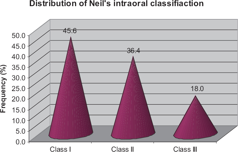
Figure 1. Distribution of tongue position of recruited subjects according to Neils’s intraoral classification
The left and right intraoral tongue position of study subjects was also assessed using special intraoral instrumentation and summarized in Table 4 and also depicted in Figure 2. On the left, 116 (23.2%) subjects intraoral were found at 10–15mm, 168 (33.6%) subjects at 16–20mm, and 216 (43.2%) subjects at 21–24mm, whereas in the right it were 104 (20.8%), 176 (35.2%), and 220 (44.0%), respectively. The tongue position increases with an increase in distance and at 16–20 and 21–24mm, the frequency was 1.6% and 0.8% higher, respectively, on the right than left, whereas at 10–15mm it was 2.4% higher in the left than in the right. Comparing the frequency distribution of left and right intraoral positions at three different levels, the χ2 test showed a similar distribution of left and right intraoral positions (χ2 = 0.88, P = 0.645), that is did not differ significantly.
Table 4. Left and right intraoral distribution of recruited subjects assessed using special intraoral instrumentation
| Distance (mm) | Left (n = 500) (%) | Right (n = 500) (%) | χ2 value | P value |
|---|---|---|---|---|
| 10-15 | 116 (23.2) | 104 (20.8) | 0.88 | 0.645 |
| 16-20 | 168 (33.6) | 176 (35.2) | ||
| 21-24 | 216 (43.2) | 220 (44.0) |
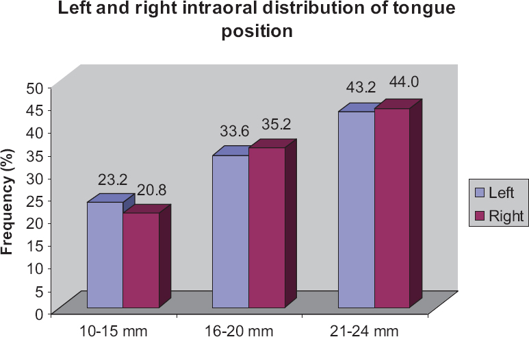
Figure 2. Left and right intraoral distribution of tongue position of study subjects assessed using special intraoral instrumentation
The distolingual area of the sulcus plays a crucial role in the creation of lower complete dentures. However, many
dentists are not familiar with it.[16] Both arches are unique, and the distolingual vestibule is not fully understood by many practitioners. When the denture is extended here, it resists horizontal forces and increases the border seal.[17] Neil has classified lateral throat form into three classes, and Edward Hartley Angle in 1989[18] has identified class I as large, class II as being between 1 and 3, and class III as shallow and unfavorable. Tuckfield[19] stated about a mechanical lock in the lateral throat form for mandibular denture retention. In addition, Hobkirk[20] mentioned that the lingual flange extension of the mandibular denture into the distolingual vestibule aids stability. This area’s distal extension is formed by the palatoglossal arch, styloglossus, superior constrictor of the pharynx, and mandibuloglossus muscle fibers.[21] Patients who become edentulous from dentulous and acquire complete dentures, lead to additional functional demands on the tongue.[22] The tongue provides speech, mastication, retention, and stability in complete dentures. In addition, it locates the mandibular denture by tactile sensation and also supports forces counteracting the dislodgement of the mandibular denture[23].
In our study, we saw that between 20 and 35 years, 210 people and between 36 and 50 years 290 people were present. Of those between 20 and 35 years, 95 males and 115 females were there. In 36–50 years, 150 males and 140 females were there. By mirror method Neil’s and by seeing tongue, it was recorded that class III was 90, class II was 182, and class I was 228. Then, by special instrument on the left maximum range was 216, the medium range was 168, and the least was 116. On the right side, the maximum range was 220, the medium range was 176, and the least was 104. This is also seen in study 17 where class I is more in younger patients. In addition, it was stated in study 9 that the distolingual flanges are shorter and narrower to the potential space when in normal function of muscles because lateral throat form in the mouth is recorded most precisely. Class I dominated more as mentioned by a study 22. More symmetry in class III was seen on both sides of the mouth. Asymmetry was seen in classes I and II because these are more common so more asymmetry there.
Clinically, this area is critical for mandibular denture stability and retention.
Classes I and II patients more than class III patients.
Vertical dimensions are more in patients’ mouths.
Tongue was more of classes I and II.
Lateral throat form classification by mirror method; then, tongue class and dimensions by instrument, all work together.
It is known that the method of classification of Neils method used in this technique was completely subjective and it has been used to no extent in research, hence it can create inconsistency between different investigators. Therefore, a technology should be developed that is more reliable and objective. This task requires patience and cooperation, as the position of the tongue affects the instrument’s placement. At times, the instrument may need to be adjusted in the mouth. To conclude with Feynman’s words, “[…] if you are doing an experiment, you should report everything that you think might make it invalid – not just what you know about it.” I think right.
Nil.
There are no conflicts of interest.
1. Devaki VN, Manonmani P, Balu K, Aravind RJ. Clinical management of highly resorbed mandibular ridge without fibrous tissue. J Pharm Bioall Sci 2012;4:149-52. [CrossRef] [PubMed] [PubMed Central] [Google Scholar]
2. Heo Y-R, Kim H-J, Son M-K, Chung C-H. Contour of lingual surface in lower complete denture formed by polished surface impression. J Adv Prosthodont 2016;8:472-8. [CrossRef] [PubMed] [PubMed Central] [Google Scholar]
3. Poudel S, Dhungana M, Karki R, Shrestha P. Lateral throat form classification using a customised gauge and Neil's method-a comparative study. J Chitwan Med Coll 2018;8:54-7. [CrossRef] [PubMed] [PubMed Central] [Google Scholar]
4. Zarb GA, Hobkirk J, Eckert S, Jacob R. Prosthodontic treatment for edentulous patients. dentures and implant supported prostheses. 12th ed. St. Louis:Mosby;2004. [Google Scholar]
5. Dipti SS, Vaishnav K, Patel P, Joshi R, Sharma K, Patel Z. Comparative evaluation of the lateral throat form and the border extension of mandibular complete denture in the distolingual region in Gandhi Nagar district:An in vivo study. Adv Hum Biol 2021;11:123-7. [CrossRef] [Google Scholar]
6. Verma M, Jaiswal R, Dubey B. Evaluative comparison of the ability of 3 border molding materials in recording the functional lateral throat form in completely edentulous mandibular arches - An in vivo study. GJRA 2018;7:24-6. [Google Scholar]
7. Kumar PR, Kalavathy N, Sridevi J, Deepak S, Mitha S, Sharmila MR. Lateral throat form measurement made easy using a customized gauge. Ann Dent Res 2011;1:100-4. [Google Scholar]
8. Kalavathy N, Kumar PR, Gupta S, Sridevi J, Shetty M, Sanketh AK. Lateral throat form re-classified using a customized gauge:A clinical study. J Indian Prosthodont Soc 2016;16:20-5. [CrossRef] [PubMed] [PubMed Central] [Google Scholar]
9. Foong KW, Patil PG. Fabrication of maxillary single complete denture in a patient with deranged mandibular occlusal plane:A case report. Saudi Dent J 2019;31:148-54. [CrossRef] [PubMed] [PubMed Central] [Google Scholar]
10. Parajuli PK, Shetty TB, Shenoy VK, Rodrigues SJ. Comparative evaluation of the lateral throat form and the border molding extension of mandibular complete dentures in the distolingual region among the south coastal Karnataka population:An in vivo study. Journal of Nepal Dental Association 2019;11:121-5. [Google Scholar]
11. Divya P, Dhawan P, Tandan P. Tongue and its prosthodontic implications. IJADS 2020;6:362. [Google Scholar]
12. Winkler S. Essentials of Complete Denture Prosthodontics. 3rd ed. Delhi, India:AITBS Publishers;2015. 112-42. [Google Scholar]
13. Bhupinder K, Gupta G, Sandhu N, Sandhu S, Kaur G, Gupta T. Tongue:The most disturbing element. Ann Dent Res 2012;2:44-51. [Google Scholar]
14. Martonet AL. Clinical application of concepts of functional anatomy and speech science to complete denture Prosthodontics. J Prosthet Dent 1962;12:817-34. [CrossRef] [Google Scholar]
15. Rinaldi P, Sharry J. Tongue force and fatigue in adults. J Prosthet Dent 1963;13:857-65. [CrossRef] [Google Scholar]
16. Engelmeier RL, Phoenix RD. Patient evaluation and treatment planning for the complete denture therapy. Dent Clin North Am 1996;40:1-18. [CrossRef] [PubMed] [Google Scholar]
17. Huang PS, Chou TM, Chang HP, Chen JH, Lee HE, Chen HS, et al. The proportion of 3 classes of lateral throat form. Int J Prosthodont 2007;20:640-2. [PubMed] [Google Scholar]
18. Angle EH. Classification of malocclusion. Dent Cosmos 1899;41:248-64. [Google Scholar]
19. Tuckfield WJ. The problem of mandibular denture. Aust J Dent 1952;56:36-49. [PubMed] [Google Scholar]
20. Hobkirk JA. The management of gross alveolar resorption. J Prosthet Dent 1973;29:397-404. [CrossRef] [PubMed] [Google Scholar]
21. Sharry, JJ. Complete Denture Prosthodontics. 3rd ed. New York, NY:McGraw-Hill. 1974. 372-76. [Google Scholar]
22. Sadhvi KV, Nair CK, Shetty J. Lateral throat form-design of a measuring instrument. KDJ 2010;33:18-9. [Google Scholar]
23. Devi LB, Keisam A, Singh HP. Malocclusion and occlusal traits among dental and nursing students of Seven North-East a states of India. J Oral Biol Craniofac Res 2022;12:86-9. [CrossRef] [PubMed] [PubMed Central] [Google Scholar]