1Department of Dentistry, Ruxmaniben Deepchand Gardi Medical College, Ujjain, Madhya Pradesh, India, 2Consultant Endodontist, Delhi, India
Address for correspondence: Dr. Ananya Bhargava, A-30/10, Vasant Vihar, Ujjain, Madhya Pradesh, India. E-mail: drananyaortho@gmail.com
Submitted: 27-Jul-2023
Revised: 21-Sep-2023
Accepted: 07-Nov-2023
Published: 03-Apr-2024
DOI: 10.4103/sidj.sidj_12_23
Saint Int Dent J 2023;7:34-8.
Copyright: © 2024 The Saint's International Dental Journal
Licence
This is an open access journal, and articles are distributed under the terms of the Creative Commons Attribution-NonCommercial-ShareAlike 4.0 License, which allows others to remix, tweak, and build upon the work non-commercially, as long as appropriate credit is given and the new creations are licensed under the identical terms.
Disclaimer:
This article was originally published by Wolters Kluwer Medknow Publications & Media Pvt Ltd and has now been officially transferred to Society of Dental Research & Education due to a change in publishing entity.
Background and Objectives: Dermatoglyphics is the study of fingerprints and skin patterns. During the intrauterine period, the development of dermatoglyphic patterns and dental hard tissues occurs at the same time. Dermatoglyphics has proved to be a potential tool in predicting dental anomalies. Hence, with the help of dermatoglyphics, we can explore the association between various dermatoglyphic patterns and types of malocclusion.
Methods: A case–control study consisting of 150 children aged 12–14 years was selected. Based on the type of occlusion, the participants were grouped into skeletal class I normal occlusion (controls), skeletal class II malocclusion, and skeletal class III malocclusion.The fingerprints were analyzed using the ink method. The molar relation was determined according to Angle’s classification of malocclusion. The relation of fingerprints was studied with the molar relation recorded using the study models. Dermatoglyphic parameters were examined in these subjects.
Results: In the study, it was observed that there was a notable increase in the occurrence of whorl pattern among patients with skeletal class II malocclusion (55%) and skeletal class III malocclusion (55.8%). On the other hand, in the skeletal class I group (controls), the frequency of loop pattern (66.6%) was found to be significantly higher. It was also noted that there was a slight decrease in the frequency of whorl patterns in skeletal class II patients as compared to skeletal class III patients.
Conclusion: The whorl fingerprint pattern found in the study may indicate a higher likelihood of skeletal class II and III malocclusion in the general population.
Keywords: Children, dermatoglyphics, malocclusion
Dermatoglyphics is the study of epidermal ridges and their configurations on the fingers, palms, and soles.[1] The term 'dermatoglyphics' is derived from two Greek words - 'derma', which means skin, and 'glyphae', which means carve. The term was coined by Cummins and Midlo in 1926.[2]
Dermal ridges begin to appear during the 12th week of intrauterine life and are completed by the 24th week of intrauterine life.[3,4] Thereafter, they remain constant, except for the change in their sizes. These dermal patterns and orodental structures embryologically develop during the same period. The three major patterns of fingerprints include arch, loop, and whorl patterns [Figure 1]. In the field of dentistry, the association of dermatoglyphics has been studied in precancerous and cancerous lesions in the oral cavity, dental caries, and dental anomalies such as cleft lip and palate and malocclusion.[5,6]
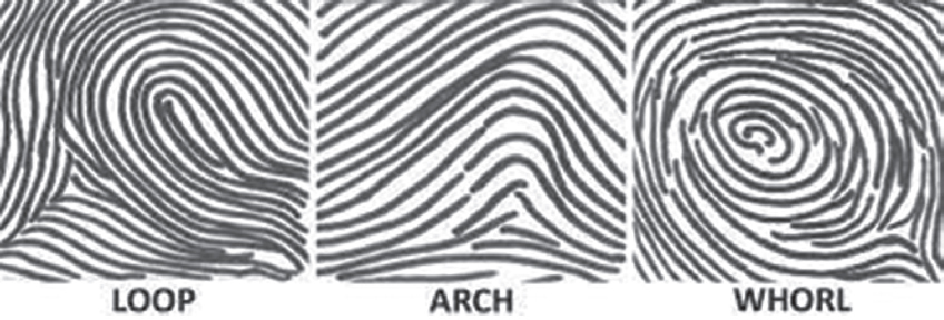
Figure 1. Fingerprint patterns
Malocclusion is a frequently reported oral condition that can be avoided, stopped, and fixed. The first step in preventing it is to be aware of the chance of developing malocclusion. Early identification of malocclusion is crucial for the best application of preventive and interceptive orthodontics.
Furthermore, relation of dermatoglyphics with dental occlusion is studied due to the fact that after the complete formation of dermal ridges, there is no change in their shape and remain in position throughout a person’s life. Thus, the present study was undertaken to ascertain the reliability of dermatoglyphics as a predictive tool for malocclusion to apply preventive and interceptive orthodontics to the high-risk groups. The purpose of the study was to study the relation of dermatoglyphics with skeletal class I normal occlusion, skeletal class II malocclusion, and skeletal class III malocclusion types in children of 12–14 years.
The study was conducted from August 1, 2021, to July 30, 2022.
This was a case–control study.
A total of 150 children from the outpatient department of dentistry, Ruxmaniben Deepchand Gardi Medical College, Ujjain, Madhya Pradesh, India, between the age group of 12–14 years were included in the study. The participants were provided with a thorough explanation of the purpose and procedures of the study, and their consent was obtained before their participation.
The researchers classified the occlusion type of 150 participants using Angle’s classification.[7] The students were then grouped based on their occlusion type.
50 skeletal class I normal occlusion (Group I/control group)
50 skeletal class II malocclusion (Group II)
350 skeletal class III malocclusion (Group III).
This was followed by recording fingerprinting for all the patients.
All permanent teeth should be present in each arch (except third molars)
No previous history of orthodontic treatment
No large coronal restorations.
Patients with a history of trauma or surgical procedures done in the orofacial region were not included.
Fingerprints were taken using standard ink method proposed by strong AM23, using blue duplicating ink (Kores India Limited, Mumbai), thick bond paper (100 g/m2).
Gloves, mouth mask, head cap, kidney tray, mouth mirror, probe, tweezer, cotton. Disinfectant, Kores India ink pad, thick white bond paper (100 g/m2), magnifying glass (6X).
The participants in the study were given instructions to wash their hands with soap and water. They were then instructed to scrub their hands thoroughly using an antiseptic lotion and allow their hands to dry. Next, the researcher guided them to press the four fingers of their right hand firmly onto an ink stamp pad against a bond paper with plate number 100 GSM. This process was then repeated with the thumb of the same hand. A hard smooth surface board was used to stabilize the paper. The same procedure was repeated for the left hand. The fingertip pattern configurations were categorized as arches, loops, and whorls [Figure 1]. Magnifying glass (6X) was used to analyze dermatoglyphic patterns. The fingerprints were analyzed qualitatively and quantitatively for arches, loops, whorls fingerprint patterns using Cummins, Midlo, and Penrose methods[8,9] [Figure 2].
Figure 2. Recorded finger and palm print of study participants by inkpad method
For recording the molar relation, alginate impressions of both the maxillary and mandibular arch were made and study models were prepared. In the study models, the molar relation was determined according to Angle’s[7] classification of malocclusion.
Normal occlusion – Normal (class I) molar relationship, teeth on line of occlusion
Class I malocclusion – Normal (class I) molar relationship teeth crowded, rotated, etc
Class II malocclusion – Lower molar distal to upper molar, relationship
Class III malocclusion – Lower molar mesial to upper molar, relationship
Using the study models relation of fingerprints with molar relationship was recorded.
Data were analyzed using IBM SPSS Statistical software for the social sciences for Windows, version 22.0 Armonk, NY, USA: IBM Corp. for the generation of descriptive and inferential statistics. The Chi-square test was used to determine the statistically significant difference among groups, and the level of statistical significance was set at P < 0.05.
Upon comparing the frequencies, we observed significant differences between the groups. The whorl pattern showed a significant increase among the skeletal class II group (55%), whereas the loop pattern was found to be significantly increased (66.6%) in the control group (P < 0.05) [Table 1].
Table 1. Comparison of fingerprint patterns among skeletal class II group and control group
| Skeletal class II group, n (%) | Control group (skeletal class I), n (%) | χ2 | P | |
|---|---|---|---|---|
| Total arch pattern | 9 (3) | 17 (5.6) | 3.4 | 0.360 |
| Total loop pattern | 126(42) | 200 (66.6) | 30.4 | 0.001* |
| Total whorl pattern | 165 (55) | 83 (27.66) | 32.8 | 0.000* |
When we compared the frequencies significant differences were noted between the groups, there was a significant increase in the whorl pattern among skeletal class III group (55.8%), whereas in the control group loop pattern (66.6%) was found to be significantly increased (P < 0.05) [Table 2].
Table 2. Comparison of fingerprint patterns among skeletal class III group and control group
| Skeletal class III group, n (%) | Control group (skeletal class I), n (%) | χ2 | P | |
|---|---|---|---|---|
| Total arch pattern | 29 (4.1) | 17 (5.6) | 8.4 | 0.079 |
| Total loop pattern | 280 (40) | 200 (66.6) | 33.2 | 0.000* |
| Total whorl pattern | 391 (55.8) | 83 (27.6) | 32.8 | 0.000* |
There was a significant increase in the whorl pattern among skeletal class II group (55%) and skeletal class III group (55.8%), whereas in skeletal class I (control) group loop pattern (66.6%)
was found to be significantly increased (P < 0.05). There is a slight decrease in the frequency of whorled patterns in skeletal class II group as compared to skeletal class III group [Table 3].
Table 3. Comparison of fingerprint patterns among skeletal class II group, skeletal class III group, and control group
| Skeletal class II group, n (%) | Skeletal class III group, n (%) | Control group (skeletal class I), n (%) | χ2 | P | |
|---|---|---|---|---|---|
| Total arch pattern | 9 (3) | 29 (4.1) | 17 (5.6) | 1.915 | 0.590 |
| Total loop pattern | 126(42) | 280 (40) | 200 (66.6) | 20.439 | 0.015* |
| Total whorl pattern | 165 (55) | 391 (55.8) | 83 (27.6) | 18.350 | 0.049* |
In the present study, there was a significant increase in the whorl pattern among skeletal class II group (55%) and skeletal class III group (55.8%). In control (skeletal class I group) loop pattern (66.6%) was found to be significantly increased.
In this study, we used the Cummins, Midlo, and Penrose ink method[8,9] to collect data. This method is cheaper, non-toxic and can be easily washed off using regular soap and water. It is ideal for collecting large-scale data. While newer digital techniques are available, they are comparatively expensive and the data obtained can be recreated or falsified. Therefore, they were not approved for study purposes.
Similar findings were observed by Eslami et al.[10] who conducted the study on 323 patients and found an increased frequency of loops and whorls and a decreased frequency of arches in all types of malocclusions.
According to a study conducted by Tikare et al.[11] and Reddy BRM et al.[12], the whorl pattern is commonly associated with skeletal class II malocclusion.
In a study conducted by Jindal et al.[13] in North India, 237 children were examined, and the results were different from earlier studies. It was found that most of the participants with any type of malocclusion had an ulnar loop pattern. In those with skeletal class III malocclusion, plain arches were the most frequently observed patterns, while in those with skeletal class II malocclusion, whorls were the most commonly observed patterns.
It should be noted that the findings presented in the current study are inconsistent with those of Rajput et al.[14] In their pilot study, they observed 24 patients with ten skeletal class I, eight skeletal class II, and six skeletal class III malocclusion. Their results indicated a higher frequency of whorls in skeletal class I malocclusion patients, and a higher frequency of loops in skeletal class II and III malocclusion patients.
The presence of a whorl fingerprint pattern may be a screening marker for malocclusion susceptibility in the general population.
Determining the genetic and environmental factors that lead to malocclusion is crucial in developing effective orthodontic treatment plans. Identifying the genetic component of the condition and a person’s proneness to it early on can help in planning preventive measures. This, in turn, can assist in selecting the right treatment modalities and ensure better outcomes for patients. Dermatoglyphic patterns can be utilized to study the genetic basis of various oral diseases such as oral cancer, oral submucous fibrosis, dental caries, periodontitis, and malocclusion. Dermatoglyphic patterns may represent the genetic makeup of an individual and therefore can be used as screening tool. Dermatoglyphics serve to strengthen the diagnostic impression of the disease and hence preventive oral health measures can be undertaken. The population at risk can be counseled and motivated to change their lifestyle, thus preventing the development of dreaded diseases in later life.
Observations should be made on a larger sample that is representative of the entire population
The quality of recorded fingerprints was dependent on the pressure and ink application, leading to improper results.
The authors would like to thank the study participants for their participation and kind cooperation throughout the study.
Nil.
There are no conflicts of interest.
1. Babu GB, Asif SM. Dermatoglyphics in dentistry:A review. Int J Contemp Dent Med Rev 2015;5;1-3. [Google Scholar]
2. Ganvir SM, Gajbhiye NY. Detection of genetic predisposition in oral squamous cell carcinoma (OSCC) and oral submucous fibrosis patients by qualitative analysis of finger and palm-print patterns:A dermatoglyphic study. Clin Cancer Invest J 2014;3:377-82. [CrossRef] [Google Scholar]
3. Shetty SS, Johnli AR, Binti NF, MdNor SN, Haron BA. Dermatoglyphics:A prediction tool for dental caries. Int J Dent Res 2016;4:30-2. [CrossRef] [Google Scholar]
4. Reddy H, Kumar P, Bari AA. Dermatoglyphics and periodontal diseases-a possible relation for early prediction. Int J Adv Res 2017;5:1332-8. [CrossRef] [Google Scholar]
5. Shetty P, Shamala A, Murali R, Yalamalli M, Kumar AV. Dermatoglyphics as a genetic marker for oral submucous fibrosis:A cross-sectional study. J Indian Assoc Public Health Dent 2016;14:41. [CrossRef] [Google Scholar]
6. Abhilash PR, Divyashree R, Patil SG, Gupta M, Chandrasekar T, Karthikeyan R. Dermatoglyphics in patients with dental caries:A study on 1250 individuals. J Contemp Dent Pract 2012;13:266-74. [CrossRef] [PubMed] [Google Scholar]
7. Angle EH. Classification of malocclusion. Dent Cosmos 1899;4:248-64. [Google Scholar]
8. Penrose LS. Fingerprints and palmistry. Lancet 1973;1:1239-42. 12. Reddy S, Prabhakar AR, Reddy VV. A dermatoglyphic predictive [CrossRef] [PubMed] [Google Scholar]
9. Penrose LS. Dermatoglyphics. Sci Am 1969;221:72-84. and comparative study of class I, class II, div. 1, div. 2 and class III [CrossRef] [PubMed] [Google Scholar]
10. Eslami N, Jahanbin A, Ezzati A, Banihashemi E, Kianifar H. . J Indian Soc Pedod Prev Dent 1997;15:13-9. Can dermatoglyphics be used as a marker for predicting future 13. Jindal G, Pandey RK, Gupta S, Sandhu M. A comparative evaluation malocclusions?Electron Physician 2016;8:1927-32. of dermatoglyphics in different classes of malocclusion. Saudi Dent [CrossRef] [PubMed] [PubMed Central] [Google Scholar]
11. Tikare S, Rajesh G, Prasad KW, Thippeswamy V, Javali SB. J 2015;27:88-92. Dermatoglyphics –A marker for malocclusion?Int Dent J 14. Rajput S, Shenoy S, Thoke B. Palmar dermatoglyphics verses 2010;60:300-4. malocclusion:A pilot study. IJRID 2014;4:48-56. [CrossRef] [PubMed] [Google Scholar]