1Department of Orthodontics and Dentofacial Orthopedics, Chhatrapati Shahu Maharaj Shikshan Sanstha’s Dental College, Aurangabad, Maharashtra, India, 2Dr. Ghunawat’s Micro-Dentistry and Advanced Dental Clinic, Aurangabad, Maharashtra, India, 3Department of Conservative Dentistry and Endodontics, MA Rangoonwala Dental College, Azam Campus, Pune, Maharashtra, India, 4Department of Orthodontics and Dentofacial Orthopedics, Chhatrapati Shahu Maharaj Shikshan Sanstha’s Dental College, Kanchanwadi, Aurangabad, Maharashtra, India
Address for correspondence: Dr. Namrata Shehare, Dr. Ghunawat’s Micro-Dentistry and Advanced Dental Clinic, Aurangabad, Maharashtra, India. E-mail: drnamratashehare@gmail.com
Submitted: 23-Dec-2022
Revised: 08-Feb-2023
Accepted: 19-May-2023
Published: 27-Jun-2023
DOI: 10.4103/sidj.sidj_20_22
Saint Int Dent J 2023;7:3-9.
Copyright: © 2023 The Saint's International Dental Journal
Licence
This is an open access journal, and articles are distributed under the terms of the Creative Commons Attribution-NonCommercial-ShareAlike 4.0 License, which allows others to remix, tweak, and build upon the work non-commercially, as long as appropriate credit is given and the new creations are licensed under the identical terms.
Disclaimer:
This article was originally published by Wolters Kluwer Medknow Publications & Media Pvt Ltd and has now been officially transferred to Society of Dental Research & Education due to a change in publishing entity.
Background: Currently, orthodontic implants have reached a peak where they are considered a dependable modality to provide temporary supplemental anchoring in orthodontic therapy. When absolute anchoring is a necessity or in cases of minimally cooperative patients, these devices can help manage skeletal anchorage. However, its failure is a serious multi-factorial issue that happens during orthodontic treatment. The stability of the mini-implant is crucial to the outcome of orthodontic intervention. Approaches to increasing the stability of the mini-implant were researched. Hence, this study was carried out to compare and contrast and clinically assess the integrity of orthodontic implants over time.
Subjects and Methods: Split mouth technique of treatment was carried out on 16 patients, i.e., one side of the mandible was considered as the experimental group (implant site irradiated with laser after placement), and the other was considered as the control side (implant site not irradiated with laser). Titanium mini-implants of the dimensions 1.5 mm diameter and 6 mm length were employed in the present study. They were positioned in the inter radicular space between the first molar and second premolar in the mandibular posterior region, 7 mm apical to the alveolar crest. During the whole process, the laser utilized was a multimode GaAs diode laser with a wavelength of 980 nm. It had 0.5–10 W output power which was adjustable with the frequency of 1–20 kHz and its main body input voltage was DC12 to further analyze the stability of the implant which in turn would aid in success assessment, the resonance frequency concept was utilized. The readings were recorded (T0) after insertion, (T1) 24 h after insertion, (T2) 2 weeks after insertion, (T3) 4 weeks after insertion, (T4) 6 weeks after insertion, and (T5) 8 weeks after insertion. The higher the implant stability quotient values the greater the stability and hence the optimal loading time.
Results: The test employed for statistical analysis was Mann–Whitney U, Kruskal Wallis, and analysis of variance test. After analysis of all the readings, it was found that low-level laser therapy has a significant role in the stability of orthodontic mini-implant.
Conclusion: The findings from this study suggest that low-level laser irradiation at the time of implant placement controls the inflammatory reaction around the implant and improves its stability.
Keywords: Anchorage, implant, laser, orthodontic implant
Orthodontic therapy subjects the teeth to a wide range of pressures and moments, including reciprocal forces. The reciprocal forces are equivalent in the magnitude and direction to the administered orthodontic forces, resulting in undesirable reciprocal movements. These undesired movements strain the anchor units, complicating and lengthening the treatment. As a result, such undesired tooth movements must be avoided. Orthodontic anchorage can be described broadly as the resistance provided by the palate, head, neck, and teeth other than those that exert unwanted reactionary tooth motions.[1-11] When absolute anchorage is a must and even a minimal space loss can affect the treatment plan supplementary aids become a necessity. As intraoral anchorage might provide only limited anchorage value, extraoral devices such as headgear or chin caps were used to reinforce the anchorage. However, esthetically unpleasant extraoral techniques have shown limited success due to patient noncompliance.[12] Orthodontic implants have gained popularity as a substitute for such extraoral appliances where there is inadequate dental reinforcement and patient concordance is uncertain. This novel method has many benefits, including simple placement, lessened patient distress, economical, quick loading, smaller diameter, adaptability in the forces to be utilized, and ease of maintenance and removal. Nonetheless, in order to improve the method’s performance level, the practitioner must efficiently manage the implants. Moreover, the success of the treatment depends on the stability of the micro-implants.[13,14] Implant failure is a major complication that occurs during orthodontic treatment. Although it is not so common, averages of 13.5% of orthodontic failure rates have been reported.[15] The failure of orthodontic micro-implants is multifactorial. Inappropriate implant placement location, as well as side, dormant period, placement guidelines, implant size, angle of an implant to the periosteum, insertion torque, extent of implant-bone interaction, amount and quality of cortical bone, severity of peri-implant inflammatory responses, thickness and mobility of soft tissue, and proximity to the root, are found to be contributing factors.[16] Peri-implantitis may be due to poor hygiene maintenance and also due to improper implant placement procedures. The overall frequency of peri-implantitis was reported to be 5%–8%.[17-20]
Measure to control peri-implantitis includes nonsurgical methods such as scaling, ultrasound, and carbon fiber curettes, curettage with antibiotics; 2% chlorhexidine or 3% hydrogen peroxide as topical antiseptics, local antibiotics such as ornidazole, metronidazole, or metronidazole with amoxicillin and surgical method is respective or regenerative techniques However, these methods take more time to cure and patient cooperation is also required.[19-21] Hence, to overcome all this, laser irradiation can be a good alternative. Low-level laser therapy (LLLT) is a type of complementary medicine treatment modality that employs low-level or near-infrared light therapy. A few basic applications of laser light therapy at the surface of the skin include reduction of pain, reduction of inflammation, prevention of damage to tissues, nerves, wounds and promotion of healing. However, it is also claimed that the laser light’s absorption affects the levels of growth hormones and inflammatory mediators, as well as cell regeneration and proliferation.
Since the inception of dental implants and lasers, significant advancement of both the techniques and the idea of combining the two have turned out to be advantageous for both the dentist and the patient. Laser therapy has been shown to have a stimulatory effect on bone or bone nodule formation in vivo or in vitro via increased expression of insulin-like growth factor (IGF) and bone morphogenetic proteins (BMPs) therefore it may enhance the stability of orthodontic mini-implant via peri-implant bone formation, which is stimulated by IGF-I and/BMPs.[22,23] Carbon dioxide, argon, holmium, and Nd: YAG, diode wavelengths fall under the category of soft-tissue lasers, whilst Er: Yttrium scandium-gallium-garnet and Erbium: YAG (Er: YAG) are considered to be hard tissue lasers.[22] After taking into consideration, the several applications of laser irradiation on implant dentistry, this study was undertaken to clinically investigate the stimulatory effect of laser irradiation on the stability of orthodontic mini-implants.
An institutional ethics committee approved this prospective two-group experimental study. Split mouth treatment was performed on 16 patients; 18–22 years old with healthy periodontium, good oral hygiene, and whose treatment plan included implant placement between mandibular 1st molar and 2nd premolar was chosen for this study from the department with patients’ informed consent. To eliminate the errors that could compromise the success of implant placement and the quality of the implant stability quotient (ISQ) reading and a stent was made out of 0.019 × 0.025” stainless steel for implant implantation. The stent’s entire length (from the bracket location) was limited to 9 mm, and its loop was 4 mm in diameter. The micro-implant was positioned 7 mm above the alveolar crest. The mesial and distal horizontal legs were ligated to the premolar bracket and molar tube respectively [Figure 1] after stent engagement, the area of the micro implant insertion was cleaned with povidone-iodine. A topical anesthetic was applied. With full retraction of the soft tissue, 0.5 ml of anesthetic solution (Lignocaine with adrenaline) was infiltrated in the mucosa. The insertion site was then evaluated with a probe to check if the area was anesthetized. This also serves as a visual marker for inserting the micro implant.
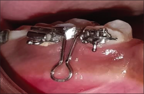
Figure 1: Stent engaged into premolar bracket and molar tube
Next, the titanium grade V self-drilling micro-implants of diameter 1.5 mm and length 6 mm[24] [Figures 2 and 3] were inserted in such a way as to obtain maximum insertion depth and an angulation of 90°. An intraoral periapical radiograph was taken to ensure that the micro implant angulation is satisfactory and to confirm that the adjacent roots were not injured [Figure 4].
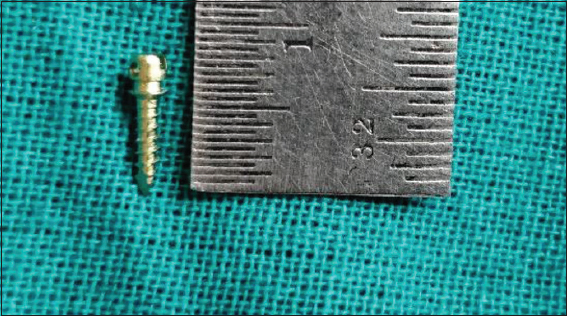
Figure 2: Micro-implant of length 6 mm and diameter 1.5 mm
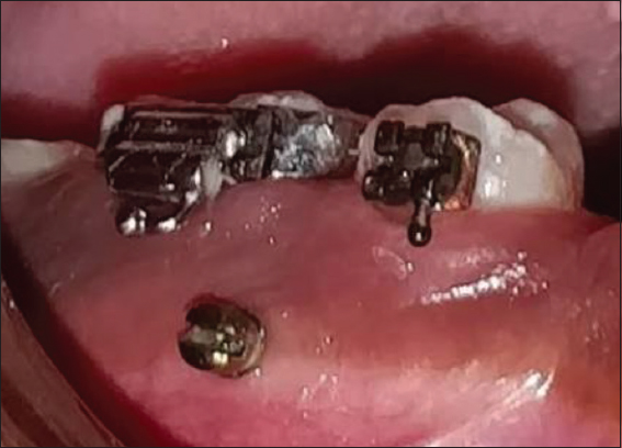
Figure 3: Micro-implant placed between 45 and 46
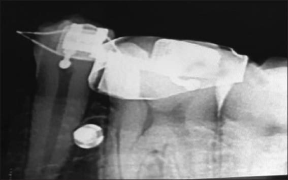
Figure 4: IOPA after micro- implant insertion. IOPA: Intraoral periapical radiograph
The irradiation site was selected by blind sampling. Sixteen envelopes, each containing eight letters RIGHT and eight letters LEFT, were prepared and kept by the nurse (who was not involved in the study). Each candidate voluntarily chose a sealed envelope, gave it to the nurse, and the nurse opened the envelope. He then informed the operator that the laser used in the procedure was a multimode GaAs diode laser with a wavelength of 980 nm. The output power was 0.5-10W, adjustable in the frequency range of 1-20KHz, and the body input voltage was DC12V~19V.
In the experimental group, implant site was irradiated with soft-tissue laser after placement, and in the control group, implant site was not irradiated with soft-tissue laser [Figure 5].
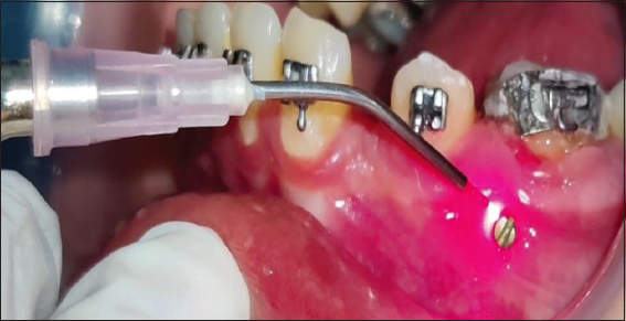
Figure 5: LLLI around the implant. LLLI: Low level laser irradiation
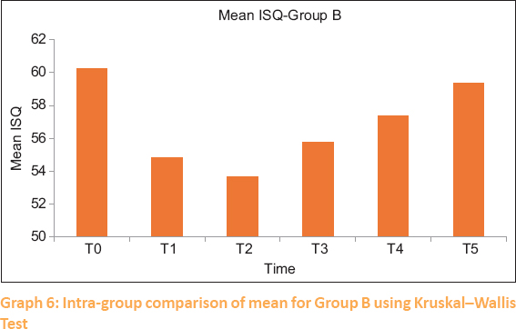
Resonance frequency analysis was used to gauge the implant’s stability. The principle for RFA assessment is the detection of the mini magnet embedded in the aluminum housing of an implant head, known as a smart peg [Figure 6]. The main unit of assessment is the resonance frequency emitted by the magnet. Because no Smart Pegs available are suitable for the orthodontic mini-implants a customized connector will be used for attachment between the two. The readings were recorded(T0) after insertion, (T1) 24 hours after insertion, (T2) 2 weeks after insertion, (T3) 4 weeks after insertion, (T4) 6 weeks after insertion, (T5) 8 weeks after insertion. Mean values were calculated for all six (3 in mesiodistal and 3 in occlusogingival directions) readings and that will be the overall ISQ value for each micro implant at each time [Figures 7-9]. The higher the ISQ values the greater will be the stability and hence the optimal loading time.
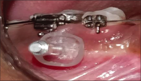
Figure 6: Smart Peg and customized connector engaged with the micro-implant head
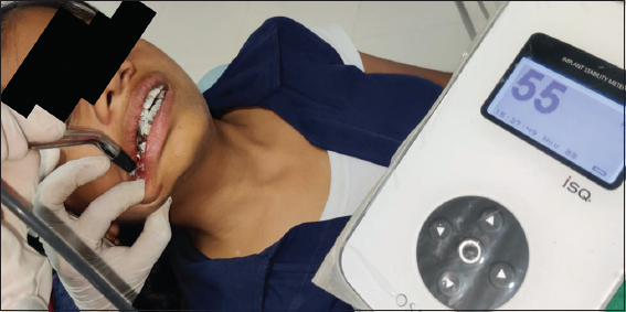
Figure 7: ISQ reading in occluso-gingival and mesio-distal direction Implant Stability Quotient ISQ (No-55)
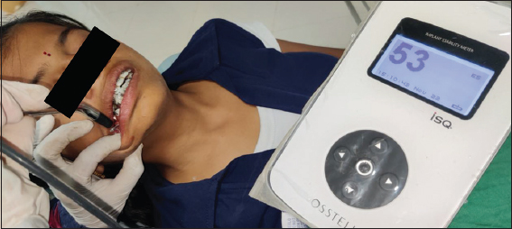
Figure 8: ISQ reading in occluso-gingival and mesio-distal direction Implant Stability Quotient ISQ (No-53)
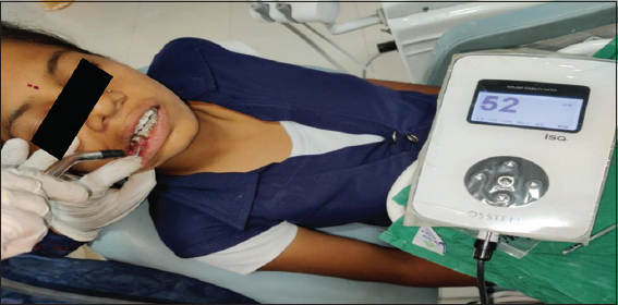
Figure 9: ISQ reading in occluso-gingival and mesio-distal direction Implant Stability Quotient ISQ (No-52)
Data were coded, transferred and analyzed on SPSS version 19 (Version 19, IBM SPSS Inc., Chicago, IL, USA). The test employed for statistical analysis was Mann-Whitney U, Kruskal Wallis and Anova test in which P value less than 0.05 was considered statistically significant.
The comparison of the stability measured in the two directions: The micro-implants have to be stable enough to sustain the forces loaded in all the directions. Thus, Mann–Whitney U-test was used to compare the stability in two perpendicular directions, i.e., Mesio-distal (D1) and occluso-gingival (D2), and then mean of all this were taken. The mean of the ISQ values was compared for each time interval (T0, T1, T2, T3, T4, and T5) in both the groups and there was no statistically significant difference found between the two directions at all-time intervals [Tables 1, 2 and Graph 1, 2].
Table 1: Comparison of the primary stability measured in the two directions for Group A by Mann–Whitney U-test
| Time interval | Mean stability measured in direction 1 | Mean stability measured in direction 2 | P | Result |
|---|---|---|---|---|
| T0 | 60.729 | 59.79 | 0.624 | Nonsignificant |
| T1 | 56.354 | 54.63 | 0.396 | Nonsignificant |
| T2 | 58.563 | 57.77 | 0.734 | Nonsignificant |
| T3 | 59.813 | 59.94 | 0.895 | Nonsignificant |
| T4 | 62.021 | 62.40 | 0.836 | Nonsignificant |
| T5 | 64.271 | 64.58 | 0.91 | Nonsignificant |
Table 2: Comparison of the primary stability measured in the two directions for Group B by Mann–Whitney U-test
| Time interval | Mean stability measured in direction A | Mean stability measured in direction B | P | Result |
|---|---|---|---|---|
| T0 | 60.31 | 60.31 | 0.624 | Nonsignificant |
| T1 | 55.00 | 55.19 | 0.396 | Nonsignificant |
| T2 | 53.75 | 54.06 | 0.734 | Nonsignificant |
| T3 | 55.63 | 56.63 | 0.895 | Nonsignificant |
| T4 | 57.69 | 58.19 | 0.836 | Nonsignificant |
| T5 | 58.88 | 60.44 | 0.91 | Nonsignificant |
Graph 1: Comparison of the primary stability measured in the two directions for Group A by Mann–Whitney U test
Graph 2: Comparison of the primary stability measured in the two directions for Group B by Mann–Whitney U test
ANOVA test was used to compare the two groups at different time intervals [Tables 3, 4 and Graphs 3, 4]. When comparing the two groups at time T0 and T1, the results were not significant. Whereas at T2, T3, T4, and T5 time intervals, the results were found to be significant.
Table 3: Multiple comparison of implant stability quotient in Group A by ANOVA
| Subset for alpha=0.05 | ||||
|---|---|---|---|---|
| 1 | 2 | 3 | 4 | |
| T1 | 55.49 | |||
| T2 | 58.17 | |||
| T3 | 59.88 | 59.88 | ||
| T0 | 60.26 | 60.26 | ||
| T4 | 62.21 | 62.21 | ||
| T5 | 64.43 | |||
Table 4: Multiple comparison of implant stability quotient in Group B by ANOVA
| Subset for alpha=0.05 | |||
|---|---|---|---|
| 1 | 2 | 3 | |
| T2 | 53.66 | ||
| T1 | 54.85 | 54.85 | |
| T3 | 55.75 | 55.75 | |
| T4 | 57.39 | 57.39 | |
| T5 | 59.38 | ||
| T0 | 60.26 | ||
Graph 3: Multiple comparison of ISQ in Group A by ANOVA. ISQ: Implant stability quotient
Graph 4: Multiple comparison of ISQ in Group B by ANOVA. ISQ: Implant stability quotient
Kruskal−Wallis test was used for intragroup comparison [Table 5, Graph 5 and 6]. In this stability was fluctuating at different time intervals in both the groups. The values are lowest at T2 in the control group and then further go on increasing till highest at T5 in experimental group it goes on increasing and crosses its baseline stability.
Table 5: Intra-group comparison of mean for Group A and Group B using Kruskal–Wallis test
| Time interval | Group A | Group B |
|---|---|---|
| T0 | 60.26 | 60.26 |
| T1 | 55.46 | 54.85 |
| T2 | 58.17 | 53.66 |
| T3 | 59.88 | 55.75 |
| T4 | 62.21 | 57.39 |
| T5 | 64.23 | 59.38 |
| P | 0.00* | 0.00* |
| Result | Significant | Significant |
Graph 5: Intra-group comparison of mean for Group A using Kruskal–Wallis test
Graph 6: Intra-group comparison of mean for Group B using Kruskal–Wallis Test
The outcome of this investigation demonstrates how the implant’s primary stability changes over time. It is better at the time of insertion, however as the inflammation sets in, the stability of the implant decreases after 24 h. This was seen in both the groups examined. In the experimental group, the stability then slowly started increasing from 2 weeks of duration to 8 weeks of duration. At 8 weeks, the stability was found to exceed the baseline line (at the time of insertion) values. In the control group, the stability declined from 24 h to 2 weeks, when the lowest ISQ values were recorded. Then, however, the stability slowly increased from the 4th week to 8 weeks’ duration. However, in the control group, the ISQ values even at 8 weeks of duration remained below the baseline values. These findings and differences between the two groups may be because of the duration of inflammatory response within the tissues adjacent to the implant. Implant placement leads to micro trauma to the tissues which leads to an inflammatory reaction. In the control group, this response is seen to last for 2 weeks of duration, whereas in the experimental group, the response lasted only for 24 h. Thus, we can report that laser irradiation has been shown to promote this treatment by reducing inflammation and edema, inducing analgesia, decreasing the inflammatory response, and accelerating tissue recovery. Furthermore, it was found that there was no significant difference in ISQ values when measured in two different directions. These findings are in agreement with those of Zita Gomes et al.,[24] Maluf et al.,[25] Khadra[26] Pinto et al.,[27] and Osman et al.[28] who have all concluded that LLLT can promote bone healing and bone mineralization and thus accelerates implant healing in the bone as well as a laser is capable of increasing the stability of self-threading orthodontic mini-implants. However, it is in dissent with the results of studies done by RPB Lobato et al.,[29] and Abohabib et al.[30] in which LLLT did not influence mini-implant stability.
The findings of this study suggest that laser irradiation at the time of implant placement controls the inflammatory reaction around the implant and improves its stability. We also recommend immediate loading or loading after 2 weeks as the implant gains its maximum stability at this time and there are lesser chances of failure.
Nil.
There are no conflicts of interest.
1. Feldmann I, Bondemark L. Orthodontic anchorage:A systematic review. Angle Orthod 2006;76:493 501. [PubMed] [Google Scholar]
2. Proffit WR. Biomechanics and mechanics. In:Proffit WR, editor. Contemporary Orthodontics. 3rd ed., Sec. IV. St Louis, Mo:CV Mosby;2000:308 11. [Google Scholar]
3. Nanda R, Kuhlberg A. Nand R. Biomechanics in clinical orthodontics. In:Biomechanical basis of extraction space closure. Philadelphia:Saunders;1997. 156-87. [Google Scholar]
4. Melsen B, Bosch C. Different approaches to anchorage:A survey and an evaluation. Angle Orthod 1997;67:23 30. [PubMed] [Google Scholar]
5. Diedrich P. Different orthodontic anchorage systems. A critical examination. Fortschr Kieferorthop 1993;54:156 71. [PubMed] [Google Scholar]
6. Roberts WE, Nelson CL, Goodacre CJ. Rigid implant anchorage to close a mandibular first molar extraction site. J Clin Orthod 1994;28:693 704. [PubMed] [Google Scholar]
7. Odman J, Lekholm U, Jemt T, Thilander B. Osseointegrated implants as orthodontic anchorage in the treatment of partially edentulous adult patients. Eur J Orthod 1994;16:187 201. [CrossRef] [PubMed] [Google Scholar]
8. Wehrbein H, Feifel H, Diedrich P. Palatal implant anchorage reinforcement of posterior teeth:A prospective study. Am J Orthod Dentofacial Orthop 1999;116:678 86. [CrossRef] [PubMed] [Google Scholar]
9. Costa A, Raffainl M, Melsen B. Miniscrews as orthodontic anchorage:A preliminary report. Int J Adult Orthodon Orthognath Surg 1998;13:201 9. [PubMed] [Google Scholar]
10. Block MS, Hoffman DR. A new device for absolute anchorage for orthodontics. Am J Orthod Dentofacial Orthop 1995;107:251 8. [CrossRef] [PubMed] [Google Scholar]
11. Melsen B, Costa A. Immediate loading of implants used for orthodontic anchorage. Clin Orthod Res 2000;3:23 8. [CrossRef] [PubMed] [Google Scholar]
12. Chen CH, Chang CS, Hsieh CH, Tseng YC, Shen YS, Huang IY, et al. The use of microimplants in orthodontic anchorage. J Oral Maxillofac Surg 2006;64:1209 13. [CrossRef] [PubMed] [Google Scholar]
13. Papadopoulos MA, Tarawneh F. The use of miniscrew implants for temporary skeletal anchorage in orthodontics:A comprehensive review. Oral Surg Oral Med Oral Pathol Oral Radiol Endod 2007;103:e6 15. [PubMed] [Google Scholar]
14. Morais LS, Serra GG, Muller CA, Andrade LR, Palermo EF, Elias CN, et al. Titanium alloy mini-implants for orthodontic anchorage:Immediate loading and metal ion release. Acta Biomater 2007;3:331 9. [CrossRef] [PubMed] [Google Scholar]
15. Papageorgiou SN, Zogakis IP, Papadopoulos MA. Failure rates and associated risk factors of orthodontic miniscrew implants:A meta analysis. Am J Orthod Dentofacial Orthop 2012;142:577 95.e7. [PubMed] [Google Scholar]
16. Baek SH, Kim BM, Kyung SH, Lim JK, Kim YH. Success rate and risk factors associated with mini-implants reinstalled in the maxilla. Angle Orthod 2008;78:895 901. [CrossRef] [PubMed] [Google Scholar]
17. Prathapachandran J, Suresh N. Management of peri-implantitis. Dent Res J (Isfahan) 2012;9:516 21. [CrossRef] [PubMed] [PubMed Central] [Google Scholar]
18. Schwarz F, Derks J, Monje A, Wang HL. Peri-implantitis. J Clin Periodontol 2018;45 Suppl 20:S246 66. [PubMed] [PubMed Central] [Google Scholar]
19. Mahato N, Wu X, Wang L. Management of peri-implantitis:A systematic review, 2010 2015. Springerplus 2016;5:105. [CrossRef] [PubMed] [PubMed Central] [Google Scholar]
20. Smeets R, Henningsen A, Jung O, Heiland M, Hammächer C, Stein JM. Definition, etiology, prevention and treatment of peri-implantitis –A review. Head Face Med 2014;10:34. [CrossRef] [PubMed] [PubMed Central] [Google Scholar]
21. Muthukuru M, Zainvi A, Esplugues EO, Flemmig TF. Non-surgical therapy for the management of peri-implantitis:A systematic review. Clin Oral Implants Res 2012;23 Suppl 6:77 83. [PubMed] [Google Scholar]
22. Pendyala C, Tiwari RV, Dixit H, Augustine V, Baruah Q, Baruah K, et al. LASERS and its applications in dentistry A contemporary apprise on LASERS and its applications in dentistry. Int J Oral Health Med Res 2017;4:47 51. [Google Scholar]
23. Omasa S, Motoyoshi M, Arai Y, Ejima KI, Shimizu N. Low-level laser therapy enhances the stability of orthodontic mini-implants via bone formation related to BMP-2 expression in a rat model. Photomed Laser Surg 2012;30:255-6. [CrossRef] [PubMed] [Google Scholar]
24. Zita Gomes R, de Vasconcelos MR, Lopes Guerra IM, de Almeida RA, de Campos Felino AC. Implant stability in the posterior maxilla:A controlled clinical trial. Biomed Res Int 2017;2017:6825213 [CrossRef] [PubMed] [PubMed Central] [Google Scholar]
25. Maluf AP, Maluf RP, da Rocha Brito C, França FM, de Brito RB. Mechanical evaluation of the influence of low-level laser therapy in secondary stability of implants in mice shinbones. Lasers Med Sci 2010;25:693-8. [CrossRef] [PubMed] [Google Scholar]
26. Khadra M. The effect of low level laser irradiation on implant-tissue interaction. In vivo and in vitro studies. Swed Dent J Suppl 2005;(172):1-63. [PubMed] [Google Scholar]
27. Pinto MR, dos Santos RL, Pithon MM, de Souza Araújo MT, Braga JP, Nojima LI. Influence of low-intensity laser therapy on the stability of orthodontic mini-implants:a study in rabbits. Oral Surg Oral Med Oral Pathol Oral Radiol 2013;115:e26-30. [PubMed] [Google Scholar]
28. Osman A, Moneim AA, El Harouni N, Shokry M. Long-term evaluation of the effect of low-level laser therapy on orthodontic miniscrew stability and peri-implant gingival condition:A randomized clinical trial. J World Federation Orthod 2017;6:109-14. [CrossRef] [Google Scholar]
29. Lobato RP, Kinalski MD, Martins TM, Agostini BA, Bergoli CD, Dos Santos MB. Influence of low-level laser therapy on implant stability in implants placed in fresh extraction sockets:A randomized clinical trial. Clin Implant Dent Relat Res 2020;22:261-9. [CrossRef] [PubMed] [Google Scholar]
30. Abohabib AM, Fayed MM, Labib AH. Effects of low-intensity laser therapy on the stability of orthodontic mini-implants:a randomised controlled clinical trial. J Orthod 2018;45:149-56. [CrossRef] [PubMed] [Google Scholar]