Apexification of the mineral trioxide aggregate in nonvital immature anterior teeth with and without platelet-rich plasma: A preliminary clinical study

Navin Mishra1, Isha Narang2, Md. Jawed Iqbal3, Priyankar Singh4, Nimmi Singh5, Ashish Pranab6

1Department of Conservative Dentistry and Endodontics, Indira Gandhi Institute of Medical Sciences (IGIMS), Patna, Bihar, India, 2Department of Dentistry, Civil Hospital, Gurgaon, Haryana, India, 3Department of Oral and Maxillofacial Surgery, IGIMS, Patna, Bihar, India, 4Department of Cranio-Maxillofacial Surgery, IGIMS, Patna, Bihar, India, 5Department of Oral Medicine and Radiology, IGIMS, Patna, Bihar, India, 6Department of Dentistry, IGIMS, Patna, Bihar, India


Address for correspondence: Dr. Navin Mishra, Department of Conservative Dentistry and Endodontics Indira Gandhi Institute of Medical Sciences (IGIMS), Bihar, India. E-mail: mishranavinendo2023@gmail.com


Submitted: 27-Apr-2023

Accepted in Revised Form: 08-Jun-2023

Published: 27-Jun-2023

DOI: 10.4103/sidj.sidj_7_23

Saint Int Dent J 2023;7:20-26.

Copyright: © 2023 The Saint's International Dental Journal

Licence

This is an open access journal, and articles are distributed under the terms of the Creative Commons Attribution-NonCommercial-ShareAlike 4.0 License, which allows others to remix, tweak, and build upon the work non-commercially, as long as appropriate credit is given and the new creations are licensed under the identical terms.

Disclaimer:

This article was originally published by Wolters Kluwer Medknow Publications & Media Pvt Ltd and has now been officially transferred to Society of Dental Research & Education due to a change in publishing entity.


ABSTRACT

Introduction: Treatment of permanent teeth with weak root dentin, exposed apex, necrotic pulp, periapical disease, and stunted root growth present a significant challenge to the practicing conservative dentistry. Necrotic immature permanent teeth with open apices are arduous to seal by thermo-plasticized or lateral condensation methods. Apexification methods have traditionally used a variety of materials including calcium hydroxide, biodentine, and mineral trioxide aggregate (MTA). Calcium hydroxide apexification takes a long time to develop an apical barrier and contains large pores, resulting in inadequate sealing ability and a bad outcome. MTA is a biomimetic and bio-inductive substance that has been employed to construct a single-step apical barrier in immature open apices with and without periapical pathology. Endodontic in this 21st century uses regenerative procedures in a nonvital, immature tooth with wide-open apices but it is also a very lengthy procedure and poor patient compliance due to slow biologic healing and long-term follow-up of the patients. Platelet-rich plasma (PRP) contains various growth factors such as platelet-derived growth factors and transforming growth factors and has been proposed as a potentially ideal scaffold for accelerated wound healing.

Aims and Objectives: To evaluate and compare the clinical and radiographic outcome of MTA apexification with and without PRP in periapical healing kinetics in nonvital, immature teeth.

Materials and Methods: An analysis was performed on patient with immature teeth, open-apex, maxillary front nonvital that were randomly divided into two groups of eighteen patients (eighteen roots permanent upper anterior) each: Group I in which MTA apexification was performed without PRP and Group II consisting of 18 roots where MTA apexification were carried out on a collagen sponge supplemented with PRP.

Results: The cases were followed up at 6 months and 12 months after treatment, both clinically and radiographically, by two independent assessors. Periapical healing (PAH) was good in 6–18 roots, and in Group I-11 roots showed excellent healing. In Group II-17 roots showed excellent results. Case number 18 and case number 9 were lost to follow-up due to migration to groups I and II, respectively. Apical closure was 100% in Group II and 77% in Group I.

Conclusions: Single-step MTA apexification with PRP is a conservative and effective method for inducing PAH and is time-saving in managing nonvital, immature teeth with an open apex.

Keywords: Apexification, collagen, mineral trioxide aggregate, open apex, platelet-rich plasma


INTRODUCTION

Tooth fracture accounts for 4%–5% of all injuries to the human body produced by either direct or indirect injury.[1] Due to their position in the oral cavity, anterior teeth (maxillary central and lateral incisors) are more prone to trauma and are particularly common among young children (45% in boys and 55% in girls),[2] leading to tooth displacement, fracture, or loss due to increased outside activities. Fractures that involve enamel (74.8%) or enamel with dentin without pulp exposure (11.7%) are characterized as simple fractures and can be repaired with various restorative procedures.[2] However, in cases of complicated tooth fractures including tooth pulp, root canal therapy must be undertaken.

The nonsurgical endodontic management of mature teeth has shown a favorable outcome rate of 95% in teeth diagnosed with irreversible pulpitis[3] and 85% in necrotic cases.[3] However, necrotic immature permanent teeth caused either by caries or trauma, offer a prognosis due to thin dentinal walls that are prone to fracture.[4-6] Moreover, open apices are difficult to seal either by thermo-plasticized or lateral condensation methods. Open apices are difficult to seal either by thermo-plasticized or lateral condensation methods. It is reported that 30% of these teeth fracture during or after such treatment.[7] Various materials have been used in apexification procedure such as calcium hydroxide, biodentin, and mineral trioxide aggregate (MTA)[7] Calcium hydroxide apexification takes a lot of time for the formation of apical barrier and has many pores which results in inadequate sealing ability and contribute to a poor outcome. MTA create an artificial apical barrier to allow optimal filling of root canal obturating material avoiding over-extrusion and has been regarded as an ideal material for perforation repair.[7,8] The chemical composition of MTA was determined by Torabinejad and Parirokh[9] developed at Loma Linda University, in the 1990s[9] introduced in dental literature in 1993 and has been approved by the Food and Drug Administration to be used in the United States in 1997 and became commercially available as ProRoot MTA.[5,10] The material consists of fine hydrophilic particles, and the main components are tricalcium silicate, tricalcium aluminate, tricalcium oxide and silicate oxide, and bismuth oxide (4:1).[11] Plethora of studies has demonstrated its excellent sealing ability and biocompatibility.[6] It is an ideal material for treating root perforation as it is nontoxic, nonabsorbable, and radiopaque and has antimicrobial properties.[7] The repair capacity of MTA can, in turn, be attributed to its antimicrobial properties due to its high pH (12.5). Regenerative endodontics can be defined as biologically based procedures designed to replace damaged structures, including dentin and root structure, as well as cells of the pulp-dentin complex.[7] The most studied and successful approach of regenerative endodontics is revascularization which is done to re-establish the vitality in a nonvital, immature tooth with wide-open, often blunderbuss apical opening to allow repair and regeneration of tissues in the form of thickening of lateral dentinal walls and elongation of the fragile, underdeveloped root. Hargreaves et al.[11] have identified three components contributing to the success of this procedure. To aid the ingrowth of new tissues into the canal space, various scaffolds material have been recommended such as synthetic polymers polylactic acid and polyglycolic acid, and natural polymers such as blood clot and collagen. Revascularization research has studied collagen as a natural scaffold in the canal space.[11,12]

Platelet-rich plasma (PRP) was first term used was “PRP” by Kingsley in 1954,[12] “Fibrin glue” was introduced by Matras in 1970,[13] called it “gelatin platelet-gel” foam in 1979, platelet-derived wound healing factors Knighton et al. in 1986,[14] first introduced in Dentistry by Whitman et al.[15] in their 1997 article entitled “Platelet gel”: Choukroun et al.[16] developed another form of PC in France which was labeled as “PRF” in year 2000, Mishra et al.[17] proposed another classification which was limited to “PRP” in year 2012 and finally Mourão et al.[18] in year 2015 gave a detailed technical note on the preparation of injectable-PRF. The authors believed that “improved wound healing can be expected through activation of the platelets within the gel and the subsequent release of growth factors.” Therefore, MTA apexification as a single-step apical barrier with PRP was used in this study, which can induce healing kinetics of the periapex and treatment can be completed in a few sittings, improving patient compliance.

Aims and objectives

Aim

  • To evaluate and compare the outcome of MTA apexification with and without PRP in periapical healing kinetics in nonvital, immature teeth.

Objectives

  • To evaluate the efficacy of MTA apexification without PRP in the healing of Periapex

  • To evaluate the efficacy of MTA apexification with PRP in the healing of Periapex

  • To compare the results of MTA apexification with and without the use of PRP in healing kinetics in immature, nonvital teeth.

MATERIALS AND METHODS

The Department of Conservative Dentistry and Endodontics at the Post Graduate Institute of Dental Education and Research at the Indira Gandhi Institute of Medical Sciences in Patna, Bihar, India, was where this study was carried out. The study was conducted in accordance with the Declaration of Ethical Criteria for Medical Research Involving Human Participants in line with the Declaration of Helsinki, after independent peer review and approval by the Institutional Ethical Committee, before commencement. Following a thorough explanation of the treatment procedure in accordance with the patient information sheet, written informed consent was acquired from the patient (or parent, if the patient was younger than 14 years of age). The treatment outcome based on clinical and radiographic criteria was assessed by calibrated examiners. Patients of both groups were recalled for follow-up examinations after the first intervention at 6 and 12 months for the 1st year, and every year thereafter until the end of the study.

Inclusion criteria

  • Patients having maxillary anterior nonvital, immature teeth with open apex/apices with or without radiographic evidence of periapical lesion

  • Involving only occlusal surface, should not be tender to percussion

  • Ideal periodontal probing depth of 2–3 mm

  • The absence of furcation radiolucencies and periapical pathology were included in the present study

  • Radiographic evidence of deep caries proximal to the pulp chamber with no evidence of periodontal ligament thickening/widening.

Exclusion criteria

  • History of any systemic conditions such as diabetes, blood dyscrasias, patients on immunosuppressants, patient with bleeding disorder, platelet count <150,000/mm, 3 Asthmatic which could compromise the healing response

  • Root Fracture and symptomatic teeth (history of spontaneous pain) and nonrestorable teeth

  • Radiographic evidence of periapical changes or pathology

  • Negative response to pulp sensibility testing

  • Tooth not conducive for rubber dam isolation

  • History of the previous restoration in the tooth under consideration for pulp capping

  • Periodontally compromised teeth and patients with unsatisfactory oral hygiene maintenance and

  • Pregnant and lactating females

  • Patient not willing to participate in the study.

Treatment methodology

Patient’s medical, dental history, demographic, and socioeconomic data were collected. Intraoral examination was performed by a principle investigator. Pulpal and periradicular status was assessed through percussion, palpation, thermal, and electric pulp tests (Diagnostic unit; Sybron, Orange, CA). Periapical radiographic examination was performed using Rinn XCP devices (Rinn Corp, Elgin, IL) with Radio Visio Graphy (Vatech, Villa India, New Delhi, India).

Sample size calculation

The sample size was calculated using a free online available software G*Power (latest ver. 3.1.9.7; Heinrich-Heine-Universität Düsseldorf, Düsseldorf, Germany) for accessing Periapical healing (PAH) after 1 year. The calculation used an equivalence limit of 0.5, a standard deviation of 0.73, an alpha level of 0.05, an 85% power test, and a two-tailed hypothesis. In addition to it, in the present study, the sample size was also increased by 18% to compensate for any drop-out. This resulted in 18 roots each comprising of total 36 nonvital, immature, maxillary incisors and all were recruited for the study. Each patient was asked to select a chit from a box randomly representing the two groups with treatment protocols. The patients were categorized into two groups having eight carious roots each: Group I in which MTA Angelus (Angelus Indústria de Produtos Odontológicos S/A, Brazil) apexification was done without PRP into the canal and Group II had 8 roots where MTA apexification was supplemented with PRP carried on a collagen sponge (Metrogene – Septodont, France).

Group I: (Mineral trioxide aggregate apexification)

The procedure of MTA Apexification was carried out as follows: Under Rubber dam isolation, access preparation was done in necrotic immature permanent teeth of all the 36 subjects. Canals were copiously irrigated with 2.5% sodium hypochlorite, and minimal instrumentation was done to prevent the weakening of the lateral dentinal walls. Triple antibiotic paste (TAP) was placed as an inter-appointment medicament in the dried canals, and the coronal access was sealed with intermediate restorative material for 4 weeks. During the visit of patients after 4 weeks under rubber dam isolation, TAP was removed from the canal using irrigation with 2.5% sodium hypochlorite. Canals were dried and following the procedure was carried out [Figure 1a-c].

thumb

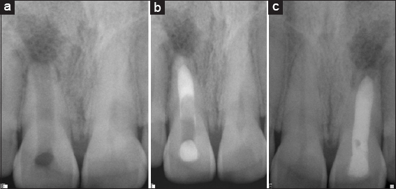
Figure 1: (a) Preoperative, (b) Mineral trioxide aggregate apexification, (c) Follow up 1 year


In Group I, MTA was placed in the canal using messing gun to form the 3–4 mm of apical plug. It was obturated using gutta-percha and AH Plus sealer. The tooth was permanently restored with adhesive restoration in the same visit.

Group II: (Mineral trioxide aggregate apexification + platelet-rich plasma on a collagen sponge)

Under Rubber dam isolation, access preparation was done in necrotic immature permanent upper anterior of 18 patients (18 roots). In the cases subjected to revascularization, local anesthesia without a vasoconstrictor was administered at the first visit, with preparation of an access cavity, working length instrumentation, irrigation with 1.5%–2.5% sodium hypochlorite and 17% ethylenediamine tetraacetic acid, Canals were copiously irrigated with 2.5% sodium hypochlorite, and minimal instrumentation was done to prevent the weakening of the lateral dentinal walls. The TAP was placed as an inter-appointment medicament in the dried canals, and the coronal access was sealed with intermediate restorative material for 4 weeks. Moreover, patients were recalled after 4 weeks of treatment. In Group II PRP + collagen was introduced as a scaffold and pushed toward the apical area using endodontic pluggers and then MTA apexification was done using messing gun to form the 3–4 mm of the apical plug. It was obturated using gutta-percha and AH Plus sealer. The tooth was permanently restored with adhesive restoration during the same visit [Figure 2a-c].

thumb

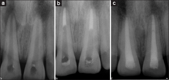
Figure 2: (a) Preoperative, (b) Mineral trioxide aggregate + Platelet rich plasma, (c) 1 year follow-up


Platelet-rich plasma preparation

It was done in a simple, tabletop laboratory centrifugation machine (Lab Care, LB-DCM8 India). 8.5 mL of whole blood was drawn by venipuncture of the antecubital vein. It was then collected in a 10 mL sterile glass tube with an anticoagulant (Acid Citrate Dextrose) in it. After 1st centrifugation at 2400 rpm for 10 min (soft spin), three separate layers were seen in the tube. After that, the PRP, soaked on a collagen sponge was introduced into the root canal and taken to the apical part of the canal and MTA apical plug was placed in the same appointment. In both the experimental groups, resin-modified glass ionomer cement was placed extending 3–4 mm in the canal. The access cavity was sealed with composite (Clearfil Majesty, Kuraray Medical Inc., Tokyo, Japan) during the same visit. Patients were kept on the follow-up of 6 and 18 months. Both clinical and radiographic evaluation was done at each follow-up visit. Clinically relief of symptoms such as pain, swelling, and resolution of sinus was evaluated and on radiograph healing of periapical lesion was noted.

RESULTS

The cases were followed up at 6 months and 12 months’ posttreatment both clinically and radiographically by two independent evaluators who were blinded about the treatment groups. The clinical evaluation considered relief of symptoms such as pain, absence of swelling, and resolution of sinus. Radiographic evaluation included PAH. Clinically both groups showed excellent results (subjective evaluation). The pain, swelling, intra-oral sinus, etc., had resolved completely in all the cases in both groups. X-rays were scored either as satisfactory (+), good (++), or excellent (+++) for all the above parameters subjectively by consensus of the two evaluators, which is presented in Table 1. Statistical comparison between Group I (MTA Apexification) and For Group II (MTA Apexification + PRP on a collagen sponge), P < 0.05 was considered to be statistically significant. Of 18 roots of PAH were good in 6 roots and 11 roots showed excellent healing in Group 1 [Table 1]. In Group II 17 cases showed excellent results [Table 2]. Case no 18 and case no 9 in Group I and II, respectively, were lost in follow-up due to migration. Apical closure was 100% in Group II and 77% in Group I.

Table 1: Intra-oral peri apical radiographic (IOPAR) observations Group I

Group I Tooth number Periapical healing MTA apexification Apical closure
Case 1 11 +++ +++
Case 2 11 +++ +++
Case 3 21 +++ +++
Case 4 11 +++ +++
Case 5 21 +++ +++
Case 6 11 ++ +++
Case 7 21 ++ ++
Case 8 11 +++ +++
Case 9 21 +++ +++
Case 10 21 ++ ++
Case 11 11 +++ +++
Case 12 21 +++ +++
Case 13 21 ++ ++
Case 14 11 +++ +++
Case 15 11 +++ +++
Case 16 21 +++ +++
Case 17 11 +++ +++
Case 18 21 No follow up No follow up

++: Good, +++: Excellent, MTA: Mineral trioxide aggregate


Table 2: Intra-oral peri apical radiographic (IOPAR) observations Group II

Group II Tooth number Periapical healing MTA + PRP Apical closure
Case 1 11 +++ +++
Case 2 11 +++ +++
Case 3 21 +++ +++
Case 4 21 +++ +++
Case 5 11 +++ +++
Case 6 21 +++ +++
Case 7 21 +++ +++
Case 8 11 +++ +++
Case 9 11 No follow up No follow up
Case 10 21 +++ +++
Case 11 21 +++ +++
Case 12 21 +++ +++
Case 13 11 +++ +++
Case 14 21 +++ +++
Case 15 11 +++ +++
Case 16 11 +++ +++
Case 17 21 +++ +++
Case 18 11 +++ +++

For statistical simplicity, satisfactory score was denoted by 1(+), good by 2(++) and excellent by 3(+++). The data was then analyzed by Chi-square test and P<0.05 was considered to be statistically significant. MTA: Mineral trioxide aggregate, PRP: Platelet rich plasma


DISCUSSION

This pilot clinical study documented the effectiveness of MTA apexification with PRP serves as a successful scaffold for regenerative endodontic treatment in nonvital, immature, anterior teeth, and speeds up the healing kinetics, saves time, and has a better patient compliance rate.[19] Conventionally, apexification with calcium hydroxide was done but it has its own disadvantages, including prolonged treatment time (6–24 months for barrier formation), patient compliance for attending the recalls, and increased risk of tooth fracture after dressing with the material for extended periods. Calcium hydroxide being hygroscopic[19] absorbs moisture from root dentin making it brittle. Its proteolytic properties[20,21] are known to alter the collagen of the ground substance of dentin adversely affecting the strength of the dentin. Both these effects make the tooth brittle and susceptible to fracture. Although the open apex might be “closed” by a calcific barrier, apexification does not promote the continued development of the root (i.e. maturogenesis). This barrier formed is often porous and not continuous. Open apex can be managed by single sitting MTA a barrier or biodentin. Periapical surgery with retrograde seal is another approach for the successful management of such teeth. However, it is an invasive procedure with its accompanying shortcomings, including the possibility of surgical complications, increased cost of treatment, and possible psychological distress, especially in children, and can also lead to a compromised crown: Root ratio in a tooth already weakened as a result of immature root development. Recently a technique based on regenerative principles termed “revascularization” or “revitalization” has been found successful in inducing maturogenesis (or apexogenesis) in cases of immature, nonvital teeth but it has major disadvantages like entire canal might be calcified making it difficult in the retreatment and In case, requiring post and core as the final restorative treatment plan, revascularization is contraindicated.[21]

Akgun et al.[22] showed that TAP when used as an intracanal medicament in immature necrotic teeth can help promote further development of the pulp-dentin complex. Hargreaves et al.[23] in their retrospective review article showed that TAP when used as an intracanal medicament in immature necrotic teeth can help promote further development of the pulp-dentin complex. Hence, in the present study, the disinfection was achieved by 5.25% of sodium hypochlorite irrigation with minimum instrumentation and by use of TAP. The formulation as suggested by Sato et al.[24] was used in the present study. Regeneration approaches[25] offer advantages over apexification, because they allow greater maturation of root in both length and thickness thanks to regenerated vital tissue and PRP can be used as a scaffold in PAH and revascularization[26] as mentioned by Hargreaves KM, et al.[11] PRP is a volume of autologous plasma that has supra physiologic platelet concentration and was introduced in dental science by Whitman et al.[15] in 1997. It is a concentrated suspension of growth factors that have been demonstrated to induce the healing and regeneration of tissues. It works via the degranulation of alpha granules that contain different growth factors that stimulate bone and soft tissue healing.[16] This present study of MTA apexification with PRP on Collagen was done because of the following proven benefits PRP helps in PAH because of the jump start osteogenesis, improves trabecular bone density, is osteoconductive, accelerates endothelial, epithelial, and epidermal regeneration and stimulates angiogenesis and collagen synthesis. Hence, the present study used PRP before MTA apexification and we achieved 100% success.

PRP can easily be procured from the patient’s own blood chair-side in the operatory. It requires a minimum quantity of blood (just 5 mL) and the machine is relatively inexpensive. As it is autologous, there is no chance of antigenic reaction. As PRP itself has an anti-inflammatory property as explained above, it does not cause any adverse inflammatory response. In the present study, collagen (Metrogene, Septodont, France) was used as a carrier for PRP. Collagen has similar chemical and biological characteristics as compared to natural tissue with low antigenicity. In the present study metronidazole containing collagen (Metrogene) was used as a carrier which has many advantages. (a) It is present in the form of a sponge and hence easy to carry PRP into the root canal. It contains resorbable collagen fibers with 4.5 mg Metronidazole which is an antimicrobial. (b) It has sustained antimicrobial activity and covers the most of diverse, polymicrobial endodontic flora from necrotic-infected root canals. (c) It is also a bio-inductive material and can improve the healing kinetics of periapical reasons. In the present study, MTA apexification was done after PRP and it showed 100% success in Healing and Periapical closure. The third important prerequisite for a successful outcome for such cases is a tight coronal seal, as coronal leakage and percolation may facilitate the invasion of canal space by bacteria and can result in treatment failure.[3] MTA has been regarded as an ideal material for apexification procedures and perforation repair.[4] The repair capacity of MTA can in turn be attributed to its antimicrobial properties due to its initial pH of 10.2, which rises to 12.5 3 h after mixing.[27] These characteristics of MTA promote the growth of cementum and the formation of new bone.[28,29]

Hence, utilizing the benefit of MTA, apexification after PRP with collagen was done and the coronal part with resin-modified glass ionomer cement and Composite was used to obtain a 3D seal. This present study achieved 100% success rate in group II when both groups were compared in PAH and apical closure in 1 year.

CONCLUSIONS

This study evaluated and compared the outcome of MTA apexification with and without PRP for apexification in nonvital, immature teeth. Thirty-six cases comprising of 34 nonvital, immature, maxillary incisors were recruited for the study as 2 subjects 1 in each group were lost up due to migration. The patients were distributed into two groups randomly: Group I in which MTA apexification was done without PRP and Group II where MTA apexification was done with PRP on a Collagen sponge. An intraoral radiograph was taken for baseline record to be compared with follow-up radiographs at 6 months and 12 months. Both clinical and radiographic evaluation was done at each follow-up visits by two independent evaluators who were blinded regarding the treatment protocol. Clinically relief of symptoms such as pain, swelling, and resolution of the sinus was evaluated and on radiograph healing of periapical lesion was evaluated. Clinically both the groups showed excellent results. The pain, swelling, and intra-oral sinus had resolved completely all the cases in both the groups. Out of 36 cases, PAH was rated as good in 4 cases and the remaining 13 cases showed excellent response in Group 1 while in Group 2, apical closure and PAH were excellent in all cases. One patient in each group was lost due to migration issues.

The present study concludes that

  • Single-step MTA apexification with PRP is a conservative and effective method for inducing PAH and is time-saving in managing nonvital, immature teeth with an open apex

  • The addition of PRP on a collagen scaffold after MTA apexification enhanced the reparative response in terms of PAH and apical closure.

Acknowledgments

This research received no grant from any funding agency in the public, commercial, or not-for-profit sectors.

Financial support and sponsorship

Nil.

Conflicts of interest

There are no conflicts of interest.

REFERENCES

1. Patnana AK, Kanchan T. Tooth fracture. In:StatPearls. Treasure Island (FL):StatPearls Publishing;2023. Available from:https://www.ncbi.nlm.nih.gov/books/NBK551650/. [Last updated on 2023 May 22]. [Google Scholar]

2. Sulieman AG, Awooda EM. Prevalence of anterior dental trauma and its associated factors among preschool children aged 3-5 years in Khartoum City, Sudan. Int J Dent 2018;2018:2135381. [PubMed] [PubMed Central] [Google Scholar]

3. Duggal M, Tong HJ, Al-Ansary M, Twati W, Day PF, Nazzal H. Interventions for the endodontic management of non-vital traumatised immature permanent anterior teeth in children and adolescents:A systematic review of the evidence and guidelines of the European academy of paediatric dentistry. Eur Arch Paediatr Dent 2017;18:139 51. [CrossRef] [PubMed] [PubMed Central] [Google Scholar]

4. Zafar K, Nazeer MR, Ghafoor R, Khan FR. Success of pulpotomy in mature permanent teeth with irreversible pulpitis:A systematic review. J Conserv Dent 2020;23:121 5. [CrossRef] [PubMed] [Google Scholar]

5. Tawil PZ, Duggan DJ, Galicia JC. Mineral trioxide aggregate (MTA):Its history, composition, and clinical applications. Compend Contin Educ Dent 2015;36:247 52. [Google Scholar]

6. Steiner JC, Dow PR, Cathey GM. Inducing root end closure of nonvital permanent teeth. J Dent Child 1968;35:47 54. [Google Scholar]

7. Kumar V, Zameer M, Prasad V, Mahantesh T. Boon of MTA apexification in young permanent posterior teeth. Case Rep Dent 2014;2014:673127. [CrossRef] [PubMed] [Google Scholar]

8. Torabinejad M, Parirokh M. Mineral trioxide aggregate:A comprehensive literature review –Part II:Leakage and biocompatibility investigations. J Endod 2010;36:190 202. [CrossRef] [PubMed] [Google Scholar]

9. Parirokh M, Torabinejad M, Dummer PM. Mineral trioxide aggregate and other bioactive endodontic cements:An updated overview –Part I:Vital pulp therapy. Int Endod J 2018;51:177 205. [CrossRef] [PubMed] [Google Scholar]

10. Schmitt D, Lee J, Bogen G. Multifaceted use of proroot MTA root canal repair material. Pediatr Dent 2001;23:326 30. [Google Scholar]

11. Hargreaves KM, Diogenes A, Teixeira FB. Treatment options:Biological basis of regenerative endodontic procedures. J Endod 2013;39:S30 43. [PubMed] [PubMed Central] [Google Scholar]

12. Kingsley CS. Blood coagulation;evidence of an antagonist to factor VI in platelet rich human plasma. Nature 1954;173:723 4. [CrossRef] [PubMed] [Google Scholar]

13. Matras H. Effect of various fibrin preparations on reimplantations in the rat skin. Osterr Z Stomatol 1970;67:338 59. [PubMed] [PubMed Central] [Google Scholar]

14. Knighton DR, Ciresi KF, Fiegel VD, Austin LL, Butler EL. Classification and treatment of chronic nonhealing wounds. Successful treatment with autologous platelet-derived wound healing factors (PDWHF). Ann Surg 1986;204:322 30. [CrossRef] [PubMed] [Google Scholar]

15. Whitman DH, Berry RL, Green DM. Platelet gel:An autologous alternative to fibrin glue with applications in oral and maxillofacial surgery. J Oral Maxillofac Surg 1997;55:1294-9. [CrossRef] [PubMed] [Google Scholar]

16. Choukroun J, Adda F, Schoeffer C, Vervelle A. PRF:An opportunity in perio implantology. Implantodontie 2000;42:55 62. [PubMed] [Google Scholar]

17. Mishra A, Harmon K, Woodall J, Vieira A. Sports medicine applications of platelet rich plasma. Curr Pharm Biotechnol 2012;13:1185 95. [CrossRef] [PubMed] [PubMed] [Google Scholar]

18. Mourão CF, Valiense H, Melo ER, Mourão NB, Maia MD. Obtention of injectable platelets rich-fibrin (i-PRF) and its polymerization with bone graft:Technical note. Rev Col Bras Cir 2015;42:421 3. [CrossRef] [PubMed] [Google Scholar]

19. Alagl A, Bedi S, Hassan K, AlHumaid J. Use of platelet-rich plasma for regeneration in non-vital immature permanent teeth:Clinical and cone-beam computed tomography evaluation. J Int Med Res 2017;45:583 93. [CrossRef] [PubMed] [PubMed Central] [Google Scholar]

20. Lekovic V, Camargo PM, Weinlaender M, Vasilic N, Kenney EB. Comparison of platelet-rich plasma, bovine porous bone mineral, and guided tissue regeneration versus platelet-rich plasma and bovine porous bone mineral in the treatment of intrabony defects:A reentry study. J Periodontol 2002;73:198 205. [CrossRef] [PubMed] [Google Scholar]

21. Shobana S, Kavitha M, Srinivasan N. Efficacy of platelet rich plasma and platelet rich fibrin for direct pulp capping in adult patients with carious pulp exposure –A Randomised controlled trial. Eur Endod J 2022;7:114 21. [Google Scholar]

22. Akgun OM, Altun C, Guven G. Use of triple antibiotic paste as a disinfectant for a traumatized immature tooth with a periapical lesion:A case report. Oral Surg Oral Med Oral Pathol Oral Radiol Endod 2009;108:e62 5. [Google Scholar]

23. Hargreaves KM, Giesler T, Henry M, Wang Y. Regeneration potential of the young permanent tooth:What does the future hold?J Endod 2008;34:S51 6. [Google Scholar]

24. Sato T, Hoshino E, Uematsu H, Noda T. In vitro antimicrobial susceptibility to combinations of drugs on bacteria from carious and endodontic lesions of human deciduous teeth. Oral Microbiol Immunol 1993;8:172 6. [CrossRef] [PubMed] [Google Scholar]

25. Nazzal H, Duggal MS. Regenerative endodontics:A true paradigm shift or a bandwagon about to be derailed?Eur Arch Paediatr Dent 2017;18:3 15. [Google Scholar]

26. Jadhav GR, Shah N, Logani A. Platelet-rich plasma supplemented revascularization of an immature tooth associated with a periapical lesion in a 40 year old man. Case Rep Dent 2014;2014:479584. [CrossRef] [PubMed] [Google Scholar]

27. Mohammadi Z, Shalavi S, Soltani MK. Mineral trioxide aggregate (MTA)-like materials:an update review. Compend Contin Educ Dent 2014;35:557-61:quiz 562. [Google Scholar]

28. Murray PE, Garcia-Godoy F, Hargreaves KM. Regenerative endodontics:A review of current status and a call for action. J Endod 2007;33:377-90. [CrossRef] [PubMed] [Google Scholar]

29. Caleza-Jiménez C, Ribas-Pérez D, Biedma-Perea M, Solano-Mendoza B, Mendoza-Mendoza A. Radiographic differences observed following apexification versus revascularization in necrotic immature molars and incisors:A follow-up study of 18 teeth. Eur Arch Paediatr Dent 2022;23:381 9. [PubMed] [Google Scholar]