MATERIALS AND METHODS
This retrospective study was conducted on the institutional ethics committee approval on lateral cephalograms obtained from the archives of the Department of Oral Medicine and Radiology. This study involves the use of 100 lateral cephalograms. All the lateral cephalograms were taken by a trained radiographic technician using the Planmeca X-mind PanoD + CEPH X-ray machine in a standard manner using the same cephalogram. The midsagittal enlargement was adjusted to 100% uniformly for all the cephalograms. All linear measurements were made with inbuilt Romexis Software. Data were includes lateral cephalogram selection for the study was based on criteria of Radiograph of patient age above 18 years and clarity of sella turcica dimensions on cephalograms. Data were not includes in study were pathology of pituitary gland and cephalograms with poor quality of images and incomplete information.
Procedure of proper method
Dimension measurements
Size of sella turcica
Three linear measurements of sella turcica, i.e. length, depth and AP diameter in mid saggital plane were obtained in accordance with Silverman and Kisling method[11,12] as shown in Figure 1.
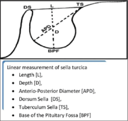
Figure 1: Three linear measurements made in sella turcica, i.e., length, depth, and AP diameter in mid saggital plane in accordance with Silverman and Kisling method. AP: Anteroposterior
Linear measurements were done on lateral cephalogram, as shown in Figure 2.

Figure 2: Linear measurement i.e., length, depth, and AP diameter is done on lateral cephalogram. AP: Anteroposterior
-
Length: The distance between the tuberculum sella (TS) to the tip of Dorsum sella [Figure 2]
-
Depth: A line perpendicular to the line drawn above the deepest point on the floor [Figure 2]
-
Anteroposterior diameter: Line drawn from the TS to the most posterior point on the posterior inner wall of the fossa [Figure 2].
RESULTS
One hundred scalp lateral cephalogram of subjects aged over 18 years were included in the present study and were treated by same orthodontist. Subjects were divided into several age groups and gender groups, of which n = 48 subjects (48.0%) were male, and n = 52 subjects (52.0%) were female. Table 1 and Chart 1 demonstrate the distribution of subjects according to age. Out of the total of 100 subjects, the age was divided into three different groups which were 11–20 years, 21–30 years, and 31–40 years. Of which, 11–20 years covered 20 subjects out of 100, while 21–30 covered 73 out of 100 subjects and 31–40 covered 7 out of 100 subjects.
Table 1: Age groups distribution of subjects
| Age groups | Subjects | Percentage |
|---|---|---|
| 11-20 | 20 | 20.0 |
| 21-30 | 73 | 73.0 |
| 31-40 | 7 | 7.0 |
| Total | 100 | 100.0 |
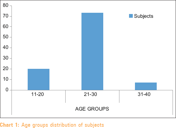
Chart 1: Age groups distribution of subjects
Table 2 and Chart 2 demonstrate the distribution of subjects according to gender. Out of the total of 100 subjects the male gender consists of 48 of a total of 100 subjects while the female consists of 53 of the total of 100 subjects. Table 3 and Chart 3 shows descriptive age comparisons of different measurements (length, depth, and anterior-posterior diameter) of Sella turcica. The mean ± standard deviation for length as a dependent variable for the 11–20 years age group is 10.7430 ± 2.29174, for the 21–30 years age group is 10.8384 ± 1.80803 and for the 31–40 years age group, is 12.3071 ± 2.42731. The total mean ± standard is 10.9221 ± 1.97226. Using one-way ANOVA, it was found that the P = 0.154, and the length is not significant with the age. The mean ± standard deviation for depth in mm as a dependent variable for the 11–20 years age group is 8.8450 ± 1.97068, for the 21–30 years age group is 8.4908 ± 1.70755 and for the 31–40 years age group is 8.6700 ± 2.24583. The total mean ± standard is 8.5742 ± 1.78666. Using one-way ANOVA, it was found that the P = 0.731, and the depth is not significant with the age. The total mean ± standard deviation for A-P diameter in mm as a dependent variable for the 11–20 years age group is 11.2715 ± 1.52919, for the 21–30 years age group is 11.5151 ± 1.66665 and for 31–40 years age group is 11.4143 ± 2.22778. The total mean ± standard is 11.4593 ± 1.66712. Using one-way ANOVA, it was found that the P = 0.846, and the anterior-posterior diameter is not significant with age.
Table 2: Gender groups distribution of subjects
| Gender groups | Subjects | Percentage |
|---|---|---|
| Female | 52 | 52.0 |
| Male | 48 | 48.0 |
| Total | 100 | 100.0 |
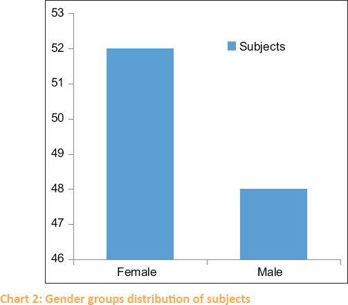
Chart 2: Gender groups distribution of subjects
Table 3: Descriptive age comparison of the measurement of sella turcica
| Age group (years) | Subjects | Measurements of sella turcica (mean±SD) | ||
|---|---|---|---|---|
| Length (mm) | Depth (mm) | A-P diameter (mm) | ||
| 11-20 | 20 | 10.7430±2.29174 | 8.8450±1.97068 | 11.2715±1.52919 |
| 21-30 | 73 | 10.8384±1.80803 | 8.4908±1.70755 | 11.5151±1.66665 |
| 31-40 | 7 | 12.3071±2.42731 | 8.6700±2.24583 | 11.4143±2.22778 |
| Total | 100 | 10.9221±1.97226 | 8.5742±1.78666 | 11.4593±1.66712 |
| ANOVA | ||||
| F | 1.909 | 0.315 | 0.167 | |
| P | 0.154 | 0.731 | 0.846 | |
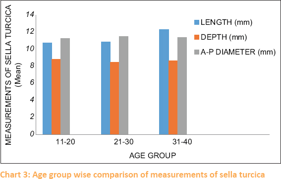
Chart 3: Age group wise comparison of measurements of sella turcica
Table 4 and Chart 4 show gender comparisons of the measurement of sella turcica. The mean ± standard deviation for length for males is 10.8010 ± 2.02251 and for females, it is 11.0338 ± 1.93766. Using the unpaired t-test, the T value, and P value were determined. The T value for length is 0.588 and the P value for length is 0.558, the length is not significant with gender. The mean ± standard deviation for depth for males is 8.1790 ± 1.71892 and for females, it is 8.9390 ± 1.78629. Using the unpaired t-test, T value and P value were determined. The T value for depth is 2.165 and the P value for depth is 0.033, the depth is significant with gender. The mean ± standard deviation for A-P diameter for males is 10.9946 ± 1.43262and for females, it is 11.8883 ± 1.76431. Using the unpaired t-test T value and P value were determined. The T value for A-P Diameter is 2.767 and the P value for the A-P diameter is 0.007, the A-P diameter is significant with gender.
Table 4: Gender comparison of the measurement of sella turcica
| Measurement of sella turcica (mean±SD) | Male | Female | Unpaired t-test | |
|---|---|---|---|---|
| t | P | |||
| Length | 10.8010±2.02251 | 11.0338±1.93766 | 0.588 | 0.558 |
| Depth | 8.1790±1.71892 | 8.9390±1.78629 | 2.165 | 0.033 |
| A-P diameter | 10.9946±10.9946 | 11.8883±1.76431 | 2.767 | 0.007 |
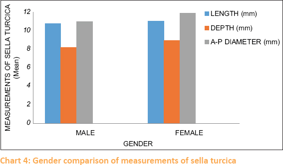
Chart 4: Gender comparison of measurements of sella turcica
DISCUSSION
The lateral cephalometric radiograph reveals many craniofacial and oral structures when imagined from the lateral aspect.[13-17] Proper analysis of these structures depends on the accurate identification and location of defined anatomical and constructed landmarks, which serve as a quantitative and qualitative measurement of lines and angles. Anatomically, sella turcica is a saddle-shaped depression in the sphenoid bone, which contains the pituitary gland in accordance with several studies done by Subasree S, Sreedevi D (2019), and Chaitanya et al. (2018), Usman et al. (2019), and Kumar et al. (2017). The exact dimensions of sella turcica are an important consideration in the diagnosis, prognosis, and treatment of diseases related to the pituitary gland and brain. The sella turcica size and morphology are different from person to person. Thus, obtaining any data in this regard will be a great help in detecting abnormalities within the anatomical area in accordance with a study done by Kumar et al. (2017).
The present study consists of a 100 sample size which is in accordance with to study done by Subasree and Sreedevi (2019) with an age range of 18 years and above and the absence of sella turcica abnormality and any pathology of sella turcica. The present study aims to find the anatomic variation of sella turcica with age. The age was divided into three distinctive groups of 11–20, 21–30, and 31–40 in accordance with a study done by Subasree and Sreedevi (2019). In the present study, on the age-wise correlation of Sella turcica length, depth, and diameter using one-way ANOVA test. it was found there was no significant relation between the age and length, depth, and A-P diameter of sella turcica. This result is in accordance with the study done by by Usman et al. (2019), and Chaitanya et al. where there was nonsignificant relation between age and Sella turcica dimensions.[4,3] This result is contrary to a study done by Subasree and Sreedevi where there was a good correlation between age and length in all age groups[1] and also to Nagaraj T. et al. (2015) where there was a significant increase in depth and anterioposterior diameter of Sella turcica as age advances.[18-21]
The present study aims to find the anatomic variation of Sella turcica with gender. In the present study, on the gender-wise correlation of Sella turcica length, depth, and diameter using t-test it was found there was a significant relationship between the gender and depth and A-P diameter of sella turcica. The mean length, depth, and A-P diameter were higher in females than in males, which was in accordance with Chauhan et al. where there was an increase in the dimensions of Sella turcica in females than in males and Anupama Deepak et al. (2018) where the mean length and depth were found to be more for females than males.[6] The study was contrary to Z. Usman et al. (2019) where the mean length, depth, and A-P diameter was higher in males than females[22,23] and Yassir et al. (2010) in Iraq population, Shah et al. (2011) in Pakistan population, Chavan et al. (2012) in Maharashtra population, Osunwoke et al. (2014) in the Nigerian, Alkofide EA (2007)where between genders, no significant difference was found in terms of length, depth, and diameter.[24,25]
CONCLUSION
This study was able to establish the basic dimensions of Sella turcica using a lateral cephalogram (length, depth, and A-P diameter). There was a statistically nonsignificant relation between the age and length, depth, and A-P diameter. There were statistically significant differences between sella turcica dimensions (depth and A-P diameter) and genders which were found in-depth and A-P diameter. Thus the linear measurement for depth and A-P Diameter will show changes in relation to males and females. And also, it was found that females tend to have greater sella turcica dimensions than those males in all linear measurements of sella turcica (length, depth, and A-P diameter).
Acknowledgment
The Corresponding author would like to thank Dr. Twinkle Patel Reader and PhD Scholar, Ahmedabad Dental College and Hospital, for his input and assistance in preparing the manuscript.
Financial support and sponsorship
Nil.
Conflicts of interest
There are no conflicts of interest.
REFERENCES
1. Subasree S, Sreedevi D. Age and gender determination using maxillary sinus and sella turcica in forensic –A lateral cephalometric study. Ind J Forensic Med To×2019;13:151 7. [CrossRef] [Google Scholar]
2. Pramod JB, Marya A, Sharma V. Role of forensic odontologist in post mortem person identification. Dent Res J (Isfahan) 2012;9:522 30. [CrossRef] [PubMed] [PubMed Central] [Google Scholar]
3. Chaitanya B, Pai KM, Chhaparwal Y. Evaluation of the effect of age, gender, and skeletal class on the dimensions of sella turcica using lateral cephalogram. Contemp Clin Dent 2018;9:195 9. [CrossRef] [PubMed] [PubMed Central] [Google Scholar]
4. Usman Z, Yunusa G, Usman J, Aliu A, Bello S. Cephalometric analysis of sella turcica for age determination from Sokoto, Nigeria –A radiological study. J Adv Med Pharm Sci 2019;21:1 7. [CrossRef] [Google Scholar]
5. Kumar TS, Govindraju P. Relationship between the morphological variation of sella turcica with age and gender:A digital radiographic study. J Indian Acad Oral Med Radiol 2017;29:164-9. [CrossRef] [Google Scholar]
6. Chauhan P, Kalra S, Mongia SM, Ali S, Anurag A. Morphometric analysisof sella turcica in North Indian Population:A radiological study. Int J Res Med Sci 2014;2:521. [CrossRef] [Google Scholar]
7. Nagaraj T, Shruthi R, James L, Keerthi I, Balraj L, Goswami RD. The size and morphology of sella turcica:A lateral cephalometric study. J Med Radiol Pathol Surg 2015;1:3 7. [CrossRef] [Google Scholar]
8. Yassir YA, Nahidh MN, Yousif HA. Size and morphology of sella turcica in Iraqi adults. Mustansiriya Dent J 2018;7:23 30. [CrossRef] [Google Scholar]
9. Chavan SR, Kathole MA, Katti AS, Herekar NG. Radiological analysis of sella turcica. Int J Recent Trends Sci Technol 2012;4:36 40. [Google Scholar]
10. Osunwoke EA, Mokwe CR, Amah-Tariah FS. Radiologic measurements of the sella turcica in an adult Nigerian population. Int J Clin Pharmacol Res 2014;4:115 7. [Google Scholar]
11. Roomaney IA, Chetty M. Sella turcica morphology in patients with genetic syndromes:A systematic review. Orthod Craniofac Res 2021;24:194 205. [CrossRef] [PubMed] [Google Scholar]
12. Muhammed FK, Abdullah AO, Rashid ZJ, Pusic T, Shbair MF, Liu Y. Morphology, incidence of bridging, and dimensions of sella turcica in different racial groups. Oral Radiol 2019;35:127 34. [CrossRef] [PubMed] [Google Scholar]
13. Sathyanarayana HP, Kailasam V, Chitharanjan AB. Sella turcica-Its importance in orthodontics and craniofacial morphology. Dent Res J (Isfahan) 2013;10:571 5. [PubMed] [Google Scholar]
14. Kucia A, Jankowski T, Siewniak M, Janiszewska-Olszowska J, Grocholewicz K, Szych Z, et al. Sella turcica anomalies on lateral cephalometric radiographs of Polish children. Dentomaxillofac Radiol 2014;43. [PubMed] [Google Scholar]
15. Magat G, Ozcan Sener S. Morphometric analysis of the sella turcica in Turkish individuals with different dentofacial skeletal patterns. Folia Morphol (Warsz) 2018;77:543 50. [CrossRef] [PubMed] [Google Scholar]
16. Tekiner H, Acer N, Kelestimur F. Sella turcica:An anatomical, endocrinological, and historical perspective. Pituitary 2015;18:575 8. [CrossRef] [PubMed] [Google Scholar]
17. Kjær I. Sella turcica morphology and the pituitary gland-a new contribution to craniofacial diagnostics based on histology and neuroradiology. Eur J Orthod 2015;37:28 36. [CrossRef] [PubMed] [Google Scholar]
18. Shah AM, Bashir U, Ilyas T. The shape and size of the sella turcica in skeletal class I, II, III in patients presenting at Islamic International Dental hospital, Islamabad. Pak Oral Dent J 2011;31:104 10. [Google Scholar]
19. Alkofide EA. The shape and size of the sella turcica in skeletal class I, class II, and class III Saudi subjects. Eur J Orthod 2007;29:457 63. [CrossRef] [PubMed] [Google Scholar]
20. Abu Ghaida JH, Mistareehi AJ, Mustafa AG, Mistarihi SM, Ghozlan HH. The normal dimensions of the sella turcica in Jordanians:A study on lateral cephalograms. Folia Morphol (Warsz) 2017;76:1 9. [CrossRef] [PubMed] [Google Scholar]
21. Tassoker M, Ozcan S. Clinical and radiological significance of sella turcica:A literature review. IOSR J Med Dent Sci 2016;15:108-3. [CrossRef] [Google Scholar]
22. Hasan HA, Alam MK, Abdullah Y, Nakano J, Yusa T, Yusof A, et al. 3D CT morphometric analysis of sella turcica in Iraqi population. J Hard Tissue Biol 2016;25:227-32. [CrossRef] [Google Scholar]
23. Hasan HA, Alam MK, Yusof A, Mizushima H, Kida A, Osuga N. Size and Morphology of Sella Turcica in Malay populations:A 3D CT Study. J Hard Tissue Biol 2016;25:313-20. [CrossRef] [Google Scholar]
24. Muhammed FK, Abdullah AO, Liu Y. A morphometric study of the sella turcica:Race, age, and gender effect. Folia Morphol (Warsz) 2020;79:318 26. [CrossRef] [PubMed] [Google Scholar]
25. Islam M, Alam MK, Yusof A, Kato I, Honda Y, Kubo K, et al. 3D CT Study of Morphological Shape and Size of Sella Turcica in Bangladeshi Population. J Hard Tissue Biol 2017;26:1-6. [CrossRef] [Google Scholar]