HEMISECTION - Salvaging the best from the rest - Case Report

Afsana Ansari1, Dipika Yadav2

1Department of Dentistry, Maharishi Deoraha Baba Autonomous State Medical College, Deoria, Uttar Pradesh, India, 2Department of Dentistry, Maa Vindhyavasini Autonomous State Medical College, Mirzapur, Uttar Pradesh, India


Address for correspondence: Dr. Dipika Yadav, Department of Dentistry, Maa Vindhyavasini Autonomous State Medical College, Mirzapur - 231 001, Uttar Pradesh, India E-mail: dipika.ydv94@gmail.com


Submitted: 19-Dec-2024,

Revised: 27-Feb-2025

Accepted: 28-Feb-2025

Published: 05-May-2025.

DOI: 10.4103/sidj.sidj_8_24

Saint Int Dent J 2024;8:52-56.

Copyright: © 2025 The Saint's International Dental Journal

Licence

This is an open access journal, and articles are distributed under the terms of the Creative Commons Attribution-NonCommercial-ShareAlike 4.0 License, which allows others to remix, tweak, and build upon the work non-commercially, as long as appropriate credit is given and the new creations are licensed under the identical terms.

Disclaimer:

This article was originally published by Wolters Kluwer Medknow Publications & Media Pvt Ltd and has now been officially transferred to Society of Dental Research & Education due to a change in publishing entity.


ABSTRACT

The term “hemisection” refers to the surgical procedure involving the sectioning of a molar tooth. This procedure entails the removal of a decayed root that may exhibit complications such as cracked roots, dental caries, or periodontal involvement in the furcation area. The primary objective of hemisection is to effectively preserve a healthy root while maintaining the integrity of the attached crown, ensuring optimal dental health and function. It is a conservative procedure to save the original tooth structure, which can act as a natural implant. The results of this procedure are predictable, and success rates are high. In this case report, hemisection was performed because the distal half of the tooth was grossly carious, and furcation was involved. The distal half of the tooth was extracted, and the remaining mesial half of the tooth was used as an abutment for the fixed partial denture. This treatment represents a conservative approach that aims to preserve as much natural tooth structure as possible. The outcomes are predictable, and the success rates in such cases are high. This case report discusses two instances of mandibular molars on which hemisection was performed, followed by restoration using fixed prostheses.

Keywords: Endo-perio lesion, Fixed Partial Denture, hemisection, mandibular molar, root caries


INTRODUCTION

One of the primary objectives of advanced dental techniques is to preserve and protect natural teeth that were once deemed irreplaceable. Nowadays, patients not only value their teeth but also prefer to keep their natural teeth instead of having them extracted whenever possible. These modern treatment methods employ a multidisciplinary approach. The terms “hemi-section” and “root amputation” refer to highly specialized procedures that are collectively categorized as “root resection.” These techniques are employed to restore the compromised function of a tooth.[1] Indications include vertical root fracture confined to a single root of a multirooted tooth or any severe destructive process that is confined to a single root, including caries, external root resorption, and trauma.[2] Periodontal, prosthodontics, and endodontic assessment for appropriate selection of cases is important.[3] The main advantage of such a treatment is the conversion of furcation-involved multirooted tooth to non-furcated single root teeth, alleviating the patient’s reluctance to teeth extraction, and providing a favorable environment for oral hygiene.[4] This case report discusses a hemisection procedure performed to preserve the endodontically treated mesial root of the mandibular left first molar while extracting the severely decayed distal root, which had furcation involvement. The remaining portion of the tooth was then utilized as an abutment for a fixed restoration.

CASE 1

A 24-year-old female patient reported to the Department of Conservative Dentistry and Endodontics, King George’s

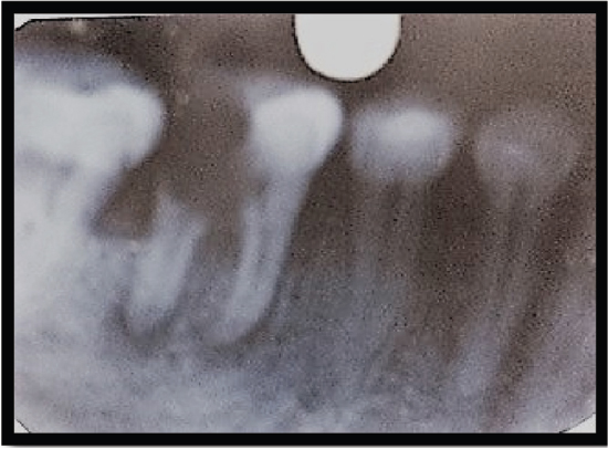
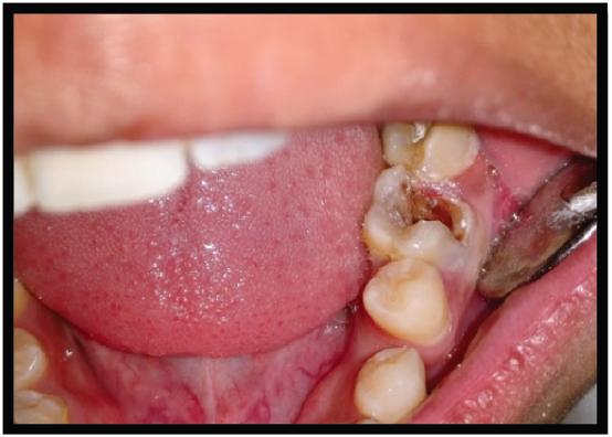
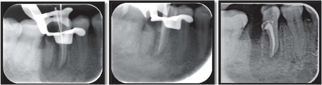
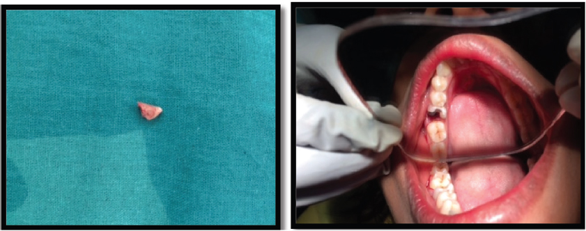
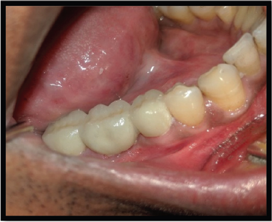
Medical College Faculty of Dental Sciences, Lucknow, with the complaint of pain in the left mandibular first molar (36) for 2-3 days. On examination, the tooth was found to be grossly carious [Figure 1]. The past medical history, as well as the past dental history of the patient, was nonsignificant. On radiographic examination, radiolucency was present in the distal root [Figure 2]. The mesial root was completely intact, but furcation was involved. It was decided that the distal root should be hemisected after the completion of endodontic therapy. Endodontic therapy was initiated with respect to the mesial root and completed in a single sitting. Canal cleaning and shaping were completed using 3% sodium hypochlorite (Septodent, France) along with 15% EDTA (Glyde, Dentsply, Tulsa, USA) and rotary file system Revo S (Micro-Mega, France). Obturation was performed by cold lateral compaction of gutta-percha [Figure 3]. Distal root hemisection was performed with a vertical cut method. The crown was cut with a tapered fissure carbide bur till the furcation was reached. After separation of both roots, the distal half was extracted [Figure 4]. The empty socket was thoroughly irrigated, and the flap was sutured. The patient was recalled for 1 month and evaluated for the healing of the extraction socket. After complete healing of the extraction socket, biomechanical preparation of the molar and premolar crown was done [Figure 5]. The reduced molar resembled a premolar in shape. A temporary bridge was seated during the healing and consolidation phase to prevent the drifting of the remaining root. Definitive restoration therapy was accomplished 6 weeks after hemisection [Figure 6]. A two-unit porcelain fused metal fixed partially replaced the distal root of the mandibular first molar and retainers on the second premolar and remaining part of the mandibular first molar. The interdental spaces are contoured so that hygiene can be accomplished with toothbrushes.

thumb

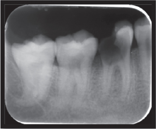
Figure 1. Grossly carious tooth

 
thumb

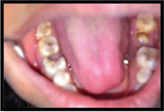
Figure 2. IOPAR showing radiolucency

 
thumb

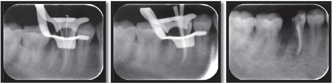
Figure 3. Steps of root canal treatment

 
thumb

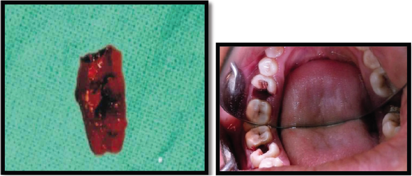
Figure 4. Hemisection of distal root

 
thumb

Figure 5. Preparation of crown

 
thumb

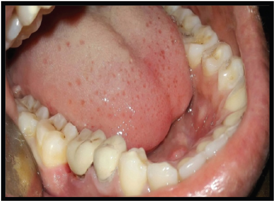
Figure 6. Post-Op 06 weeks


CASE 2

A 26-year-old female patient presented to the Faculty of Dental Sciences at King George’s Medical University, Lucknow, with the primary complaint of pain and sensitivity in the right lower posterior region persisting for the past 3 months. The pain was triggered by mastication and was relieved thereafter. A radiograph (IOPA) revealed periodontal bone loss affecting the coronal third of the distal root compared to the mesial root, and it showed periapical radiolucency at the distal root [Figure 7]. The periodontal support of the mesial root of tooth 46 was deemed adequate. Based on these findings, the diagnosis of chronic irreversible pulpitis associated with the lower right mandibular molar, along with furcation involvement, was established [Figure 8]. It was determined that the distal root should undergo hemisection subsequent to the completion of endodontic therapy for the tooth, in accordance with the methodology employed in Case 1 [Figures 9]. The hemisection of the distal root was executed using a vertical cut technique to effectuate separation. The crown was sectioned using a tapered fissure carbide bur until it reached the furcation. The procedure was thoroughly elucidated to the patient, and informed consent was duly obtained. Following the administration of local anesthesia, a mucoperiosteal flap was reflected to expose the area designated for hemisection. The hemisection procedure was performed on tooth 46 [Figure 10], after which the flapwas repositioned and sutured using 3-0 silk nonresorbable interrupted sutures. Definitive restorative therapy was conducted 6 weeks after the hemisection, which involved the placement of a three-unit porcelain fused-to-metal fixed partial denture to replace the distal root of the mandibular first molar, second premolar, and second molar [Figure 11]. At the 1-year follow-up appointment, the patient reported being asymptomatic.

thumb

Figure 7. IOPAR showing grossly carious tooth

 
thumb

Figure 8. IOPAR showing involved furcation

 
thumb

Figure 9. Steps of root canal treatment

 
thumb

Figure 10. Hemisection of distal root

 
thumb

Figure 11. Follow up


DISCUSSION

This case report presents a detailed examination and treatment of a severely damaged tooth exhibiting furcation involvement. In our case, tooth was effectively treated through hemisection, a powerful alternative to extraction that preserves natural tooth structure and promotes better long-term outcomes. The remaining roots underwent root canal therapy and were restored with suitable materials and then splinted to an adjacent tooth to prevent displacement. A fixed prosthodontic prosthesis was subsequently placed to maintain occlusal balance. The hemisection procedure allows physiologic tooth mobility of the remaining root, which is more suitable abutment for fixed partial dentures.[5] Root amputation is a useful alternative treatment to save those decayed multi-rooted teeth which have been indicated for extraction.[6] Before selecting a tooth for hemisection, the patient’s oral hygiene status and medical status should be considered, and the accessibility of root furcation for ease of operation as well as good bone support for the remaining roots should be assessed.[7] The furcation region is carefully smoothed to allow proper cleaning to prevent accumulation of food lodging.[8] Root fracture is the main cause of failure after hemisection, so occlusal modification is required to balance the occlusal forces on the remaining root.[9] Contraindications include the presence of a strong abutment tooth adjacent to the proposed hemisection, which could act as an abutment to the prosthesis.[10] Hemisection has been used successfully to retain teeth with furcation involvement. However, there are few disadvantages associated with it. As with any surgical procedure, it can cause pain and anxiety. Root surfaces that are reshaped by grinding in the furcation or at the site of hemisection are more susceptible to caries. Often, a favorable result may be negated by decay after treatment. Failure of endodontic therapy due to any reason will cause failure of the procedure[11] Furthermore, there are conflicting data about the survival of the remaining fragment (3%-38% for 10 years) and a wide range of reasons for the failure of hemisection[12] According to Buhler et al. hemisection should be considered before every molar extraction because this procedure can provide good absolute biological cost savings with good long-term success.[13] In the present case, good prognosis was observed with proper occlusion, the absence of mobility, and healthy periodontal tissues. Concurring with previous reports, hemisection is a valid treatment option for the molar teeth in a young patient, which otherwise have to be extracted due to extensive caries [Figures 9-11].

CONCLUSION

The hemisection procedure for a tooth necessitates meticulous planning and execution. It is imperative that the patient demonstrates a high level of motivation and maintains optimal oral hygiene. When these criteria are satisfied, hemisection can serve as a viable alternative to tooth extraction, followed by replacement options including conventional fixed or removable prostheses or dental implants.

Declaration of patient consent

The authors certify that they have obtained all appropriate patient consent forms. In the form, the patient(s) has/have given his/her/their consent for his/her/their images and other clinical information to be reported in the journal. The patients understand that their names and initials will not be published and due efforts will be made to conceal their identity, but anonymity cannot be guaranteed.

Financial support and sponsorship

Nil.

Conflict of interest

There are no conflict of interest.

REFERENCES

1. Chen B, Zhu Y, Lin M, Zhang Y, Li Y, Ouyang X, et al. Expert consensus on the diagnosis and therapy of endo-periodontal lesions. Int J Oral Sci 2024;16:55. [CrossRef] [PubMed] [PubMed Central] [Google Scholar]

2. Sato M, Garcia-Sanchez A, Sanchez S, Chen IP. Use of 3-dimensional-Printed guide in hemisection and autotransplantation of a fusion tooth:A case report. J Endod 2021;47:526-31. [CrossRef] [PubMed] [PubMed Central] [Google Scholar]

3. Saluja I, Kamath AK, Pradeep S, Gupta R, Duggal K. Hemisection:Partial preservation of compromised tooth. J Conserv Dent 2023;26:355-8. [CrossRef] [PubMed] [PubMed Central] [Google Scholar]

4. Khetan RR, Jr, Mahapatra J. Hemisection:A boon for the hopeless tooth. Cureus 2024;16:59967. [CrossRef] [PubMed] [PubMed Central] [Google Scholar]

5. Lin TS, Allosias JA, Halim MS, Ahmed N, Heboyan A. Restorative option for hemisected root canal treated mandibular molar:Two case reports. J Surg Case Rep 2025;2025:rjaf051. [CrossRef] [PubMed] [PubMed Central] [Google Scholar]

6. Taori P, Nikhade PP, Mahapatra J. Hemisection:A different approach from extraction. Cureus 2022;14:29410. [CrossRef] [Google Scholar]

7. Sharma S, Sharma R, Ahad A, Gupta ND, Mishra SK. Hemisection as a conservative management of grossly carious permanent mandibular first molar. J Nat Sci Biol Med 2018;9:97-9. [CrossRef] [PubMed] [PubMed Central] [Google Scholar]

8. Radke U, Kubde R, Paldiwal A. Hemisection:A window of hope for freezing tooth. Case Rep Dent 2012;2012:390874. [CrossRef] [PubMed] [PubMed Central] [Google Scholar]

9. Ganesan K, Balagangadharan M, Sengoden T, Santhi B, Vasudevan M, Dhamodharan YT. Hemisection-A Challenge for Perio-endo Lesions:A case report. J Pharm Bioallied Sci 2020;12:S631-4. [CrossRef] [PubMed] [PubMed Central] [Google Scholar]

10. Passarelli PC, Pagnoni S, Piccirillo GB, Desantis V, Benegiamo M, Liguori A, et al. Reasons for tooth extractions and related risk factors in adult patients:A cohort study. Int J Environ Res Public Health 2020;17:2575. [CrossRef] [PubMed] [PubMed Central] [Google Scholar]

11. Babaji P, Sihag T, Chaurasia VR, Senthilnathan SH. A conservative management of periodontally involved molar tooth in a young patient. J Nat Sci Biol Med 2015;6:253-5. [CrossRef] [PubMed] [PubMed Central] [Google Scholar]

12. Schwendicke F, Graetz C, Stolpe M, Dorfer CE. Retaining or replacing molars with furcation involvement:A cost- effectiveness comparison of different strategies. J Clin Periodontol 2014;41:1090-7. [CrossRef] [PubMed] [Google Scholar]

13. Hanafi L. An approach of preserving a mandibular primary second molar by a hemisection procedure:A case report with 36-month follow up. Heliyon 2022;8:10655. [CrossRef] [PubMed] [PubMed Central] [Google Scholar]