Estimation of superoxide dismutase and glutathione peroxidase in oral precancerous and cancerous lesions: A comparative assessment

Ghanshyam S. Yadav1, Devesh Tiwari2, Akhtar Husain2, Mohammad Zeeshan3, Pawan Srivastava4

1Department of Oral Medicine and Radiology, Azamgarh Dental College, Azamgarh, Uttar Pradesh, India, 2Dental Surgery Department, Hind Institute of Medical Sciences, Ataria, Sitapur, Uttar Pradesh, India, 3Department of Oral and Maxillofacial Surgery, Career Institute of Dental and Medical Sciences, Lucknow, Uttar Pradesh, India, 4Department of Dental Surgery, Era Medical College, Lucknow, Uttar Pradesh, India.

Address for correspondence: Dr. Akhtar Husain, Assistant Professor, Dental Surgery Department, Hind Institute of Medical Sciences, Atari, Sitapur - 261 303, Uttar Pradesh, India. E-mail: dr.akhtar007@gmail.com


Submitted: 07-Jan-2025,

Revised: 17-Mar-2025,

Accepted: 21-Mar-2025,

Published: 05-May-2025.

DOI: 10.4103/sidj.sidj_2_25

Saint Int Dent J 2024;8:42-47.

Copyright: © 2025 The Saint's International Dental Journal

Licence

This is an open access journal, and articles are distributed under the terms of the Creative Commons Attribution-NonCommercial-ShareAlike 4.0 License, which allows others to remix, tweak, and build upon the work non-commercially, as long as appropriate credit is given and the new creations are licensed under the identical terms.

Disclaimer:

This article was originally published by Wolters Kluwer Medknow Publications & Media Pvt Ltd and has now been officially transferred to Society of Dental Research & Education due to a change in publishing entity.


ABSTRACT

Superoxide dismutase (SOD), catalase (CAT), glutathione peroxidase (GPx), reduced glutathione (GSH), and glutathione reductase (GR) are recognized as primary enzymatic antioxidants. These antioxidants may be produced by tumor cells or generated in response to biological reactions associated with tumor growth. Excessive production of reactive oxygen species (ROS) combined with inadequate elimination can lead to a significant deficiency in antioxidant levels, resulting in an imbalance between oxidants and antioxidant defense systems. This imbalance contributes to oxidative stress (OS), which in turn can cause irreparable damage to cells and tissues, playing a critical role in the initiation, promotion, and progression of cancer. This study investigated the serum levels of superoxide dismutase (SOD) and glutathione peroxidase (GPx) in healthy individuals, patients with oral precancerous lesions, and cancer patients in order to identify potential biomarkers. The present study included 150 blood samples collected from 150 participants, of both genders, aged between 18 and 80 years. The subjects were classified into three distinct groups: Group A (control), Group B (precancerous), and Group C (cancerous). The findings indicated that participants in the oral cancer group exhibited a statistically significant reduction (P < 0.001) in the mean levels of SOD and GPx when compared to the control group, with the cancerous group demonstrating the lowest levels among all study groups. This observed imbalance in the antioxidant enzyme status may be a key factor in the pathogenesis of cancer and has the potential to serve as a biomarker and therapeutic target for mitigating malignant transformation in oral premalignant lesions and conditions.

Keywords: Antioxidants, biomarker, oral cancer, oxidative stress, premalignant


INTRODUCTION

Oral cancer is a critical form of cancer that requires urgent attention and awareness. It arises from the oral epithelium, with a global annual incidence of over 400,000 cases and an expected increase of 40%, including mortality. Principal factors contributing to the development of oral squamous cell carcinoma (OSCC) include personal behavior such as smoking, tobacco chewing, and alcohol consumption.[1-3] Oral cancer primarily develops due to socioeconomic factors, environmental conditions, and local issues like trauma or sharp teeth. Viral infections and genetic mutations in oncogenes or tumor suppressor genes also significantly contribute to oral carcinogenesis.

The human body effectively produces a range of enzymatic and non-enzymatic antioxidants that serve as vital defense mechanisms against reactive oxygen species (ROS). These unstable free radicals, including superoxide (O2) and hydroxyl radicals (OH), pose significant threats to healthy cells, compromising their structure and function, and can ultimately lead to malignancy.[4,5] Antioxidants are essential for inhibiting the formation of these harmful radicals, ensuring robust protection against oxidative stress and its damaging effects.

Superoxide dismutase (SOD), catalase (CAT), glutathione peroxidase (GPx), reduced glutathione (GSH), and glutathione reductase (GR) are the primary enzymatic antioxidants.[6] Other antioxidants include ß carotene, vitamins B complex, C, and E and the mineral selenium. These antioxidants are released by the tumor cells or the due to the body’s response to tumor growth. When the generation of the ROS exceeds the preferred levels due to excessive accumulation/reduced elimination, the consequent deficient level of antioxidants may lead to a disproportion between oxidants and antioxidant défense systems, leading to oxidative stress (OS). The induced OS results in irreparable cellular/tissue damage, which is vital in cancer initiation, promotion, and progression.[4-7]

The glutathione antioxidant system comprises key enzymes, including glutathione peroxidase (GPx), GR, and glutathione S-transferases (GST). GPx is essential for removing reactive species by reducing lipid hydroperoxides to alcohols and is mainly found in the cell membrane and cytoplasm. SOD converts superoxide radicals (O2) to hydrogen peroxide (H2O2), which GPx and catalase then turn into water. This process neutralizes reactive species, while SOD induction helps maintain GPx activity. GSH serves as a cofactor for GPx and GST in detoxifying lipid peroxides, protecting cells from free radical damage. GPx catalyzes GSH, forming oxidized glutathione (GSSG), which is then converted back to GSH by GR, ensuring a high GSH/GSSG ratio in the cell.[8-10]

To counter these consequences, the major antioxidant defense systems consisting of SOD and GPx are apparently responsible for scavenging free radicals and nascent oxygen. Therefore, considering the high prevalence of oral cancer and OSMF in India, which has not been widely studied with regard to lipid peroxidation and antioxidants, a need is felt for this study which may help in the early diagnosis and to monitor the progression of these conditions and to estimate and compare the levels of serum superoxide dismutase and glutathione peroxidase in normal individuals and oral precancerous and cancerous patients.

MATERIALS AND METHODS

The study was designed with 150 participants, aged 18 to 80. The study protocol was explained, and they were recruited for this study, all of whom provided informed consent. They were selected from the Department of Oral Medicine and Radiology at the Career Post Graduate Institute of Dental Sciences & Hospital in Lucknow and the Department of Dental Surgery at the Hind Institute of Medical Sciences in Sitapur, UP, India. The participants were classified into three groups of 50: Group A (Control), Group B (patients with precancerous lesions), and Group C (patients with cancerous lesions). Each subject was provided with a definitive diagnosis following rigorous and comprehensive clinical examinations, complemented by meticulous biochemical and histopathological investigations, ensuring the highest level of accuracy and reliability in their assessment [Table 1].

Table 1: Details of study groups

Study group Subjects as per inclusion and exclusion criteria Size
Group A Normal healthy individuals without any oral lesions, with/ without any habit (smoking and tobacco) 50
Group B Individuals with precancerous lesions, with smoking and tobacco habits 50
Group C Individuals with cancerous lesions, with smoking and tobacco habits 50
 

Inclusion criteria

Patients were clinically and histopathologically diagnosed with oral precancer and oral cancer. Normal subjects were those having tobacco-chewing habit but without any oral lesions and systemic diseases

Exclusion criteria

The exclusion criteria were as follows: patients below the age of 18 and above 80 years; patients suffering from any systemic disease, for example, diabetes, hypertension, cardiovascular disease, renal dysfunction, and liver disorder; and patients undergoing or those who had undergone the same treatment.

METHODS

Sample collection and lysate preparation

Under aseptic conditions, blood samples were obtained from each subject following an overnight fasting period. A volume of 5 milliliters of venous blood was extracted using a sterile syringe, with rigorous measures implemented to prevent hemolysis. For the analysis of superoxide dismutase and glutathione peroxidase, 3 mL of blood was placed in an EDTA vial and centrifuged at 3000 rpm for 10 min to effectively separate the plasma. The erythrocytes were washed twice using 0.9% NaCl solution at 10000 rpm for 15 min at 40 degrees Celsius. The supernatant was discarded each time, and equal volumes of chilled triple-distilled water were added to the RBC pellet. A final centrifugation at 10,000 rpm for 10 min at 4 degrees Celsius resulted in the collection of the supernatant (lysate), which was then stored at –80 degrees Celsius for subsequent analysis. For protein estimation, a 2-mL blood sample was placed in a plain vial and left to clot for 2 hours. Following this, the sample was centrifuged at 3000 rpm for 10 min to separate the serum, which was immediately utilized for total protein analysis.

Statistical analysis

Descriptive statistics were employed to present the baseline characteristics of the patients. Means and standard deviations (SD) were calculated for the data. For variables that did not follow a normal distribution, the Mann–Whitney U test and the Kruskal–Wallis test were utilized. To examine the relationships between clinical, biochemical, and demographic parameters, Pearson’s chi-square test was applied. The analysis was conducted using SPSS software (version 26.3, IBM Corp., Armonk, NY, USA). A statistical significance level of P < 0.05 was established.

RESULTS

This comparative study involved 150 participants, consisting of 136 males and 14 females, aged between 18 and 80 years. The subjects were divided into three groups: Group A (control), Group B (precancerous), and Group C (cancerous), with 50 subjects in each group based on specific inclusion and exclusion criteria. The aim was to estimate the levels of SOD and GPx in oral precancerous and cancerous lesions. The age ranges for each group were as follows: Group A ranged from 38 to 69 years, Group B from 28 to 78 years, and Group C from 26 to 72 years. The mean ages (± SE) for each group were 50.72 ± 0.89 years for Group A, 47.12 ± 1.69 years for Group B, and 49.92 ± 1.69 years for Group C, with median ages of 50 years, 45 years, and 50 years, respectively. In terms of the gender distribution, Group A had five females (10.0%) and 45 males (90.0%), Group B comprised seven females (14.0%) and 43 males (86.0%), while Group C included two females (4.0%) and 48 males (96.0%).

In this study, the SOD levels (measured in U/ml) were observed in three groups: Group A, Group B, and Group C. The SOD levels ranged from 1.23 to 15.91 for Group A, 0.74 to 5.36 for Group B, and 0.83 to 3.27 for Group C. The means (± SE) for each group was as follows: Group A had a mean of 5.57 ± 0.52, Group B had a mean of 2.70 ± 0.16, and Group C had a mean of 1.93 ± 0.09. The medians for the groups were 3.9 for Group A, 2.6 for Group B, and 1.9 for Group C. The results indicate that the mean SOD level was highest in Group A, followed by Group B, and lowest in Group C (Group A > Group B > Group C). This trend suggests that as the severity of the condition increases, the mean SOD levels decrease [Figure 1 and Table 2].

thumb

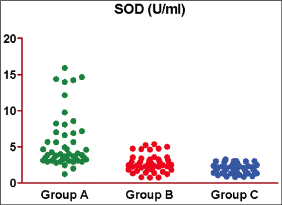
Figure 1: Scatterplot showing the observed SOD of three groups


Table 2: Basic characteristics of three groups

Basic characteristics Group A (n = 50) (%) Group B (n = 50) (%) Group C (n = 50) (%) F/χ2 P value value
Age (years)
Mean ± SE 50.72 ± 0.89 47.12 ± 1.69 49.92 ± 1.69 1.59 0.207
Sex
Female 5 (10.0) 7 (14.0) 2 (4.0)
Male 45 (90.0) 43 (86.0) 48 (96.0) 2.99 0.224
 

The mean differences in SOD between the groups were observed. The Tukey test showed a significantly different and lower SOD in both Group B (51.5%) (5.57 ± 0.52 vs. 2.70 ± 0.16, q = 8.97, P < 0.001) and Group C (65.3%) (5.57 ± 0.52 vs. 1.93 ± 0.09, q = 11.31, P < 0.001) as compared to Group A [Table 3 and Figure 2]. However, it did not differ between Group B and Group C (2.70 ± 0.16 vs. 1.93 ± 0.09, q = 2.34, P = 0.226), although it was lower (28.4%) in Group C as compared to Group B [Table 3 and Figure 3].

Table 3: SOD (U/ml) of three groups

Group Min Max Mean SE Median
Group A 1.23 15.91 5.57 0.52 3.9
Group B 0.74 5.36 2.70 0.16 2.6
Group C 0.83 3.27 1.93 0.09 1.9
 
thumb

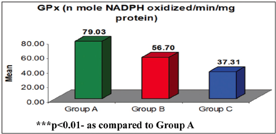
Figure 2: Mean GPx of three groups. ***P < 0.01 – as compared to Group A

 
thumb

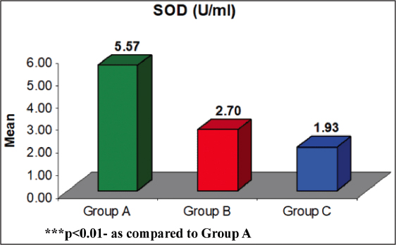
Figure 3: Mean SOD of three groups. ***P < 0.01 – as compared to Group A


The difference in the mean SOD between groups were compared by Tukey test showing significantly different SOD between Group A and Group B (q = 8.97, P < 0.001) and Group A and Group C (q = 11.31, P < 0.001) but similar between Group B and Group C (q = 2.34, P = 0.226) [Figure 4].

thumb

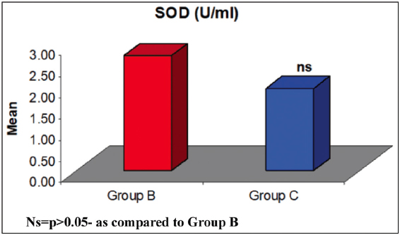
Figure 4: Comparison of mean SOD between two groups. Ns = P > 0.05 – as compared to Group B


The observed GPx (n mole NADPH oxidized/min/mg protein) of three groups is presented in Table 4 and Figure 5.

Table 4: Comparison of mean differences in SOD between the groups by Tukey test

Comparison Mean difference q value P value 95% CI (mean difference)
Group A vs. Group B 2.87 8.97 <0.001 1.80 to 3.95
Group A vs. Group C 3.62 11.31 <0.001 2.55 to 4.70
Group B vs. Group C 0.75 2.34 0.226 −0.32 to 1.82
 
thumb

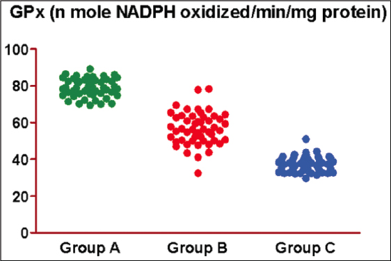
Figure 5: Scatter plot showing observed GPx of three groups


It has been observed that the GPx of Group A, Group B, and Group C ranged from 69.42–89.24, 32.51–78.39, and 29.70–51.08, respectively, with mean (± SE) 79.03 ± 0.70, 56.70 ± 1.26, and 37.31 ± 0.59, respectively, and median 78.8, 56.3, and 37.3, respectively. The mean GPx of Group A was the highest, followed by Group B, and Group C the least (Group A > Group B > Group C) In other words, as severity increases, the mean GPx decreases. It has been found that as regards the mean difference in GPx between the groups, Tukey’s test showed significantly different and lower GPx in both Group B (28.3%) (79.03 ± 0.70 vs. 56.70 ± 1.26, q = 24.90, P < 0.001) and Group C (52.8%) (79.03 ± 0.70 vs. 37.31 ± 0.59, q = 46.51, P < 0.001) as compared to Group A. Furthermore, it was also reduced significantly in Group C (34.2%) as compared to Group B (56.70 ± 1.26 vs. 37.31 ± 0.59, q = 21.61, P < 0.001) [Table 5 and Figure 2].

Table 5: GPx (n mole NADPH oxidized/min/mg protein) of three groups

Group Min Max Mean SE Median
Group A 69.42 89.24 79.03 0.70 78.8
Group B 32.51 78.39 56.70 1.26 56.3
Group C 29.70 51.08 37.31 0.59 37.3
 

The differences in mean GPx between groups were compared by Tukey’s test and showed significantly different GPx between Group A and Group B (q = 24.90, P < 0.001), Group A and Group C (q = 46.51, P < 0.001), and Group B and Group C (q = 21.61, P < 0.001) [Figures 6 and 7, and Table 6].

thumb

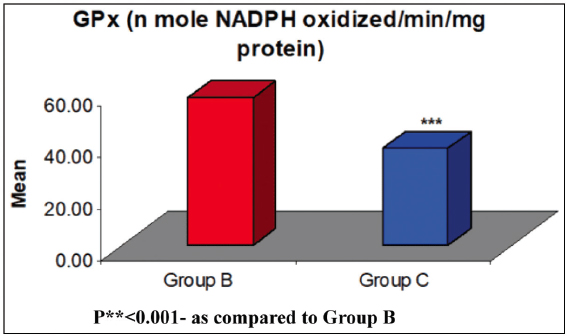
Figure 6: Comparison of mean GPx between two groups. P** < 0.001 – as compared to Group B

 
thumb

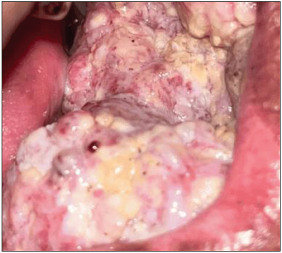
Figure 7: Clinical image of an oral cancer, included in this study


Table 6: Comparison of mean difference in GPx between the groups by Tukey’s test

Comparison Mean difference q value P value 95% CI (mean difference)
Group A vs. Group B 22.33 24.90 <0.001 19.32 to 25.34
Group A vs. Group C 41.71 46.51 <0.001 38.70 to 44.72
Group B vs. Group C 19.38 21.61 <0.001 16.37 to 22.39
 

DISCUSSION

The study cohort consisted of 91 males (91%) and nine females (9%) who exhibited behaviors that could potentially lead to malignant transformations in the body due to their high cytotoxicity, including the ability to block protective enzymes. These behaviors included tobacco use, betel nut consumption, and betel chewing. Previous studies have demonstrated that these habits have clastogenic and carcinogenic effects. The fundamental hypothesis is that free radicals damage cellular components, potentially triggering the transformation of normal cells into malignant ones. However, the extent of this damage depends on the body’s defense mechanisms, which are mediated by various cellular antioxidants. The two verified mechanisms favoring radical alteration of ROS metabolism in cancer cells are the production of huge amounts of ROS compared with non-neoplastic cells and the suppression of the antioxidant system.[3,4]

The enzyme GPx is a selenocysteine-dependent antioxidant that plays a crucial role in neutralizing hydrogen peroxide (H2O2). It relies on reduced glutathione (GSH) to function effectively. GSH provides the necessary reducing equivalents, enabling GPx to catalyze the conversion of hydrogen peroxide to water. Additionally, increased GPx activity can influence glutathione levels. GPx is essential for detoxification, particularly in low hydrogen peroxide concentrations.[10-15] Conversely, when the GPx pathway is saturated or when there is an excess of hydrogen peroxide, CAT becomes active. A recent study found that the activity of GPx was significantly reduced in erythrocyte and plasma samples obtained from patients with cancerous lesions (Group C) compared to the precancerous and normal control groups (P < 0.05). Specifically, the GPx activity was lower in the oral cancer group than in the control group, with a pooled standardized mean difference of −2.766 moles/min/g Hb at a 95% confidence interval of (–3.297 to –2.234). This increased GPx activity may be due to a heightened response to the toxic substances produced by tumor cells, serving to protect the surrounding unaffected cells.[16-19] Supporting this notion, another study indicated that elevated levels of salivary antioxidants could be a compensatory defensive reaction. Conversely, some studies suggest that the depletion of glutathione peroxidase in advanced malignancies may result from an increased utilization of antioxidants from circulation, aimed at countering excessive free radicals and resulting in oxidative damage.[6,20-22] Moreover, levels of GPx expression have been positively correlated with favorable prognoses, particularly in patients with advanced stage IV tumors, according to one reported study.

In the present study, we observed a statistically significant decrease in the levels of SOD and GPx in cancerous lesions compared to the corresponding control and precancerous lesions (P < 0.001). This finding is consistent with those of previous studies. Patients with oral leukoplakia exhibited slightly lower levels of SOD and GPx than those with oral submucous fibrosis (OSMF),[8,23-26] but this difference was not statistically significant (P > 0.05). Notably, prior literature on the comparison of antioxidant enzyme statuses between OSMF and oral leukoplakia patients is limited. In this study, the oral cancer group displayed a significant (P < 0.001) reduction in mean levels of SOD and GPx compared to the control group, showing the lowest levels among all study groups. This suggests that the decreased activity of antioxidant enzymes in oral cancer patients may stem from the depletion of the antioxidant defense system, overwhelmed by the elevated levels of lipid peroxides. The low GPx levels indicate that most cancer cell types are unable to effectively detoxify hydrogen peroxide. Additionally, alterations in the temporal pattern of thiobarbituric acid reactive substances have been linked to circadian fluctuations in antioxidant enzymes in oral cancer patients.[25,26] To establish accurate cutoff values for SOD and GPx as biomarkers for oral cancer, further research with a large sample size across multiple centers is necessary.

CONCLUSION

The current study found that levels of glutathione peroxidase (GPx) and superoxide dismutase (SOD) activity were significantly reducedincancerouslesions.Inpoorlydifferentiatedlesions, the activities of both GPx and SOD were lower compared to those of well-differentiated lesions. Additionally, in cases with larger lesion sizes (T3–T4) and lymph node involvement (N3–N4), the levels of these antioxidant enzymes were also diminished. Therefore, biological markers related to oxidative stress could be valuable for the diagnosis and treatment of oral cancer. In conclusion, the levels of antioxidant enzymes, specifically SOD and GPx, are important due to their potential role in various cancerous conditions and are essential components of the cellular antioxidant defense mechanisms.

Future directions

Further comprehensive studies involving larger sample sizes of oral submucous fibrosis (OSMF) and oral leukoplakia across various clinical stages are essential to provide clarity. Furthermore, rigorous histopathological grading and follow-up evaluations are necessary to accurately ascertain the role of these biochemical parameters in the initiation and advancement of carcinogenesis.

Disclosures

Human/animal subjects: all authors have confirmed that this study did not involve human participants/animal subjects or tissue.

Financial support and sponsorship

Nil.

Conflicts of interest

There are no conflict of interest.

REFERENCES

1. Zanaruddin SN, Yee PS, Hor SY, Kong YH, Ghani WM, Mustafa WM, et al. Common oncogenic mutations are infrequent in oral squamous cell carcinoma of Asian origin. PLoS One 2013;8:e80229. [CrossRef] [Google Scholar] [PubMed Central] [PubMed]

2. Mohideen K, Krithika C, Jeddy N, Bharathi R, Thayumanavan B, Sankari SL. Meta-analysis on risk factors of squamous cell carcinoma of the tongue in young adults. J Oral Maxillofac Pathol 2019;23:450-7. [CrossRef] [Google Scholar]

3. Mohideen K, Jeddy N, Krithika C, Faizee SH, Dhungel S, Ghosh S. Assessment of glutathione peroxidase enzyme response and total antioxidant status in oral cancer - Systematic review and metaanalysis. Cancer Rep (Hoboken) 2023;6:e1842. [CrossRef] [Google Scholar] [PubMed Central] [PubMed]

4. Liochev SI, Fridovich I. The effects of superoxide dismutase on H2O2 formation. Free Radic Biol Med 2007;42:1465-9. [CrossRef] [Google Scholar]

5. Chole RH, Patil RN, Basak A, Palandurkar K, Bhowate R. Estimation of serum malondialdehyde in oral cancer and precancer and its association with healthy individuals, gender, alcohol, and tobacco abuse. J Cancer Res Ther 2010;6:487-91. [CrossRef] [Google Scholar] [PubMed]

6. Choudhari SK, Chaudhary M, Gadbail AR, Sharma A, Tekade S. Oxidative and antioxidative mechanisms in oral cancer and precancer:a review. Oral Oncol 2014;50:10-8. [CrossRef] [Google Scholar] [PubMed]

7. Silva PVD, Troiano JA, Nakamune ACMS, Pessan JP, Antoniali C. Increased activity of the antioxidants systems modulate the oxidative stress in saliva of toddlers with early childhood caries. Arch Oral Biol 2016;70:62-6. [CrossRef] [Google Scholar] [PubMed]

8. Yang J, Lam EW, Hammad HM, Oberley TD, Oberley LW. Antioxidant enzyme levels in oral squamous cell carcinoma and normal human oral epithelium. J Oral Pathol Med 2002;31:71-7. [CrossRef] [Google Scholar] [PubMed]

9. Valko M, Rhodes CJ, Moncol J, Izakovic M, Mazur M. Free radicals, metals and antioxidants in oxidative stress-induced cancer. Chem Biol Interact 2006;160:1-40. [CrossRef] [Google Scholar]

10. Subapriya R, Kumaraguruparan R, Nagini S, Thangavelu A. Oxidant-antioxidant status in oral precancer and oral cancer patients. Toxicol Mech Methods 2003;13:77-81. [CrossRef] [Google Scholar] [PubMed]

11. FlohéL. The impact of thiol peroxidases on redox regulation. Free Radic Res 2016;50:126-42. [CrossRef] [Google Scholar] [PubMed]

12. Korde SD, Basak A, Chaudhary M, Goyal M, Vagga A. Enhanced nitrosative and oxidative stress with decreased total antioxidant capacity in patients with oral precancer and oral squamous cell carcinoma. Oncology (Huntingt) 2011;80:382-[CrossRef] [Google Scholar] [PubMed]

13. Khanna R, Thapa PB, Khanna HD, Khanna S, Khanna AK, Shukla HS. Lipid peroxidation and antioxidant enzyme status in oral carcinoma patients. Kathmandu Univ Med J (KUMJ) 2005;3:334-9. [CrossRef] [Google Scholar] [PubMed]

14. Bose SC, Singh M, Vyas P, Singh M. Plasma zinc antioxidant vitamins, glutathione levels and total antioxidant activity in oral leukoplakia. Dent Res J (Isfahan). 2012;9:158-61. [CrossRef] [Google Scholar] [PubMed Central] [PubMed]

15. Traverso N, Ricciarelli R, Nitti M, Marengo B, Furfaro AL, Pronzato MA, et al. Role of glutathione in cancer progression and chemoresistance. Oxid Med Cell Longev 2013;2013:972913. [CrossRef] [Google Scholar] [PubMed Central] [PubMed]

16. Gurudath S, Naik RM, Ganapathy KS, Guruprasad Y, Sujatha D, Pai A. Superoxide dismutase and glutathione peroxidase in oral submucous fibrosis, oral leukoplakia, and oral cancer:A comparative study. J Orofac Sci 2012;4:114-9. [CrossRef] [Google Scholar]

17. Mirnic J, Djuric M, Veljovic T, Gusic I, Katanic J, Vukoje K, et al. Evaluation of lipid peroxidation in the saliva of diabetes mellitus Type 2 patients with periodontal disease. Biomedicines 2022;10:3147. [CrossRef] [Google Scholar] [PubMed Central] [PubMed]

18. Strycharz-Dudziak M, Fołtyn S, Dworzański J, Kiełczykowska M, Malm M, Drop B, et al. Glutathione Peroxidase (GPx) and Superoxide Dismutase (SOD) in Oropharyngeal Cancer Associated with EBV and HPV Coinfection. Viruses 2020;12:1008. [CrossRef] [Google Scholar] [PubMed Central] [PubMed]

19. Srivastava KC, Austin RD, Shrivastava D, Sethupathy S, Rajesh S. A Case control study to evaluate oxidative stress in plasma samples of oral malignancy. Contemp Clin Dent 2012;3:271-6. [CrossRef] [Google Scholar] [PubMed Central] [PubMed]

20. Manasaveena V, Akula K, Sangram V. A comparative evaluation of enzymatic antioxidant levels in pre and post therapy patients with oral cancer. Int J Pharm Pharm Sci 2014;6:52-6. [CrossRef] [Google Scholar]

21. Gurudath S, Ganapathy K, D S, Pai A, Ballal S, Ml A. Estimation of superoxide dismutase and glutathione peroxidase in oral submucous fibrosis, oral leukoplakia and oral cancer--a comparative study. Asian Pac J Cancer Prev 2012;13:4409-12. [CrossRef] [Google Scholar] [PubMed]

22. de Martel C, Georges D, Bray F, Ferlay J, Clifford GM. Global burden of cancer attributable to infections in 2018:A worldwide incidence analysis. Lancet Glob Health. 2020;8:e180-90. [CrossRef] [Google Scholar] [PubMed]

23. Sharma M, Rajappa M, Kumar G, Sharma A. Oxidant-antioxidant status in Indian patients with carcinoma of posterior one-third of tongue. Cancer Biomark 2009;5:253-60. [CrossRef] [Google Scholar] [PubMed]

24. Yokoe H, Nomura H, Yamano Y, Fushimi K, Sakamoto Y, Ogawara K, et al. Characterization of intracellular superoxide dismutase alterations in premalignant and malignant lesions of the oral cavity:Correlation with lymph node metastasis. J Cancer Res Clin Oncol 2009;135:1625-33. [CrossRef] [Google Scholar] [PubMed Central] [PubMed]

25. Mohideen K, Chandrasekaran K, Dhungel S, Ghosh S. Assessment of antioxidant enzyme superoxide dismutase (SOD) in oral cancer:Systematic review and meta-analysis. Dis Markers 2024;2024:2264251. [CrossRef] [Google Scholar] [PubMed Central] [PubMed]

26. Dhanya M, Kumar Vadivel J, Umamaheswari TN, Jayaraman S. Comparative assessment of lipid peroxidase in oral cancer and oral potentially malignant disorders. Cureus 2024;16:e66474s. [PubMed] [PubMed Central] [Google Scholar] [CrossRef]