Emergency removal of traumatic foreign particles from submandibular region - A case report

Zibran Khan1, Arun Tondon2, Gargi Jadaun1, Soham Nevkar1, Priyanka Takhellambam1

1Department of Oral and Maxillofacial Surgery, Pacific Dental College and Hospital, Udaipur, Rajasthan, India, 2Department of Oral and Maxillofacial Surgery, Daswani Dental College and Hospital, Kota, Rajasthan, India


Address for correspondence: Dr. Gargi Jadaun, Department of Oral and Maxillofacial Surgery, Pacific Dental College and Hospital, Udaipur - 313 024, Rajasthan, India. E-mail: gargisingh6@gmail.com


Submitted: 04-Jun-2024

Revises: 14-Aug-2024

Accepted: 23-Aug-2024

Published: 16-Oct-2024.

DOI: 10.4103/sidj.sidj_2_24

Saint Int Dent J 2024;8:20-23.

Copyright: © 2024 The Saint's International Dental Journal

Licence

This is an open access journal, and articles are distributed under the terms of the Creative Commons Attribution-NonCommercial-ShareAlike 4.0 License, which allows others to remix, tweak, and build upon the work non-commercially, as long as appropriate credit is given and the new creations are licensed under the identical terms.

Disclaimer:

This article was originally published by Wolters Kluwer Medknow Publications & Media Pvt Ltd and has now been officially transferred to Society of Dental Research & Education due to a change in publishing entity.


ABSTRACT

Angle grinders are undeniably one of the most perilous tools utilized in industry and agriculture, with over 5000 injuries documented, and they can even be fatal if major blood vessels in the neck are ruptured and neurological damage occurs. Angle grinders can cause severe injuries to the head, neck, and face because their high-speed discs do not respect the body’s natural boundaries or structures. These injuries can be disfiguring, permanently disabling, or even fatal. To attain aesthetically pleasing outcomes, it is imperative to thoroughly cleanse the wound, excise any damaged tissue, and meticulously close the wound in layers. The article presents a case of a penetrating injury caused by using an angle grinder and discusses effective management and prevention of such injuries.

Keywords: Foreign body, injury, management, maxillofacial, penetrating<


INTRODUCTION

Penetrating neck injuries are encountered in 5-10% of all trauma cases.[1] The head, neck, and face encompass intricate and delicate organ systems, housing vascular and nervous structures, and are routinely exposed to the external environment.[2] Due to the nature of these areas, injuries are more frequently experienced than in other regions of the body. Foreign body injuries have the potential to cause immediate life-threatening and delayed complications, which can result in long-lasting impairment. Therefore, it is crucial to identify and remove foreign bodies during clinical assessments. Imaging studies play a vital role in significantly increasing the likelihood of detection and in guiding treatment planning.

Decision-making on removing foreign bodies in the maxillofacial region is complex and depends on factors like size, accessibility, and proximity to vital structures.[3] We present a case of foreign body entrapment and its management under general anesthesia.

CASE REPORT

A 45-year-old conscious and well-oriented male was presented to the emergency department with painful swelling in the left cheek and a wound dressing in the left submandibular region, which had been in place for two hours [Figure 1]. The patient exhibited positive responsiveness to verbal commands and initially received first aid treatment at a rural health facility. The patient demonstrated positive responsiveness to verbal commands and initially received first aid treatment at a rural health facility. The patient’s medical history revealed that the trauma was a result of contact with a fragment of a grinding wheel rotating at high speed. Furthermore, upon extraoral examination, a laceration measuring approximately 4 × 2 cm was identified in the left body-angle region, affecting the skin and the underlying platysma muscle. A substantial clot and green grinding disc fragments were observed within the wound. The patient exhibited limited ability to rotate his head to the left. Intraorally, no discrepancies or instability in occlusion were noted, and there was no active bleeding at the injury site. A computed tomography (CT) scan was advised to the patient. Skull base and vertebrae fracture were ruled out. It revealed multiple radiopaque foreign objects scattered all over the submandibular and the retromandibular region, with the largest piece (2.2 × 0.5 cm) presented posterior to the left ramus of the mandible inferior to the left parotid gland. It also revealed a hematoma of 7 × 5 cm in size about the left mandible body and ramus region which corroborated the clinical finding [Figure 1]. The case was taken as an emergency. Wound debridement under GA was the treatment plan decided. The incision was made by widening the existing laceration present extraorally. The hematoma was carefully removed with the help of gauze. Foreign bodies were visualized after the hematoma removal, especially the ones present in the retromandibular region. The wound was thoroughly explored, and disc fragments were removed. The larger piece was removed using a curved artery taking care not to damage any adjacent vital structures [Figure 2]. There was a presence of powder of the foreign body which was cleared out using copious saline irrigation. Closure was done in multiple layers using 3-0 vicryl sutures. Aggressive pre- and post-operative antibiotic coverage was given. Extraoral pressure bandage was given to obliterate any dead space. The post-operative period was uneventful. The patient was followed up for the next 3 months with no further complaints [Figure 3].

thumb

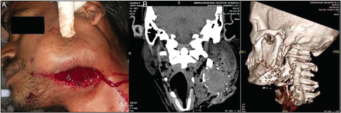
Figure 1: (A) Pre-operative picture of the patient. (B) Pre-operative CT scan

 
thumb

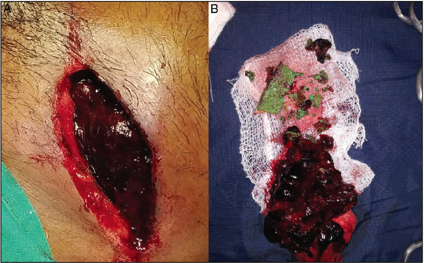
Figure 2: (A) Wound debridement. (B) The presence of hematoma and glass piece

 
thumb

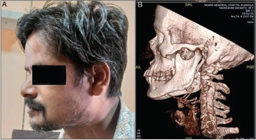
Figure 3: (A) Post-operative picture of the patient. (B) Post-operative CT scan


DISCUSSION

Foreign bodies pose potentially life-threatening risks to patients based on type, size, and location. They can result from accidents, self-inflicted injuries, iatrogenic causes, assaults, and work-related incidents.[2,4] Wood, metal, stones, broken teeth, plastics, fish bones, maggots, projectiles, and glass are the most commonly incorporated materials.[2,5,6] Penetrating facial injuries inflicted by the use of rotating angle grinders[4] are recognized sources of serious morbidity as in our case.

Penetrating injuries can occur in three horizontal anatomic zones of the neck as described in the literature.[5] In the present case, the injuries were confined to Zone II that is the space between the cricoid cartilage and the angle of the mandible. It includes the carotid and vertebral arteries. There was no damage to major vessels as well as the aerodigestive tract in this case.[5]

A detailed clinical examination assessing the wound size and severity should be carried out.[7] It demands a multidisciplinary systematic evaluation focusing on airway maintenance, bleeding control, and neurological status.[4]

Imaging studies enhance foreign body detection. Ultrasound is suitable for superficial injuries, while magnetic resonance imaging is recommended for injuries involving radiolucent bodies and excluding ferromagnetic ones.[7] Arterial angiography, contrast-enhanced CT, color Doppler ultrasonography, and magnetic resonance angiography offer vital insights into vascular injuries in penetrating neck injury cases.[5] In our case, direct CT scan was advised.

Angle grinder wounds typically mirror the cutting disc’s shape, often curvilinear but may vary with the entry angle. Tissue loss is common. The presence of disc fragments and cut material in the wound is pathognomonic for angle grinder injuries,[8] as seen in our case.

Choosing the right surgical access is crucial for planning and achieving a successful surgical outcome. Key considerations include lesion size and type, surrounding anatomy, trauma extent, patient’s health, and the surgeon’s experience and preference while selecting a surgical approach.[9]

In our case with good surgical access, surgery was completed without sacrificing major vascular and neural structures. The existing laceration was utilized to provide adequate exposure, allowing for a direct line of access to extract a deeply located grinding wheel. This approach provided us with a shorter distance to the target, which facilitated precise foreign body extraction and ultimately shortened the operative time.

Our patient was followed up for the next 3 months with no post-operative complications.

CONCLUSION

Even though facial penetrating injuries, such as in this case, can have catastrophic results, a straightforward approach through an accurate-staged sequence and critical judgment can ensure satisfactory cosmetic and functional outcomes.

Declaration of patient consent

All appropriate patient consent forms were obtained. In the form, the patient has given his consent for his images and other clinical information to be reported in the journal. The patient understand that name and initials will not be published, and due efforts will be made to conceal identity, but anonymity cannot be guaranteed.

Author contributions

Zibran Khan: Designed the study, Manuscript editing, Manuscript preparation, editing, and review. Arun Tandon: Data analysis and acquisition, Manuscript preparation. Gargi Jadaun: Definition of the intellectual content, Data acquisition and analysis. Priyanka Jadaun: Literature search, Data analysis and acquisition, Manuscript preparation.

Soham Nevkar: Literature search, Data analysis and acquisition.

Financial support and sponsorship

Nil.

Conflicts of interest

There are no conflicts of interest.

REFERENCES

1. Vishwakarma R, Joseph ST, Sharma A, Gupta RC. Endoscopic removal of sharp foreign bodies in neck using image intensified television. Int J Head Neck Surg 2010;1:113-6. [CrossRef] [Google Scholar]

2. Voss JO, Maier C, Wuster J, Beck-Broichsitter B, Ebker T, Vater J, et al. Imaging foreign bodies in head and neck trauma:A pictorial review. Insights into Imaging 2021;12:1-16. [CrossRef] [PubMed] [PubMed Central] [Google Scholar]

3. Al-Ahmady HH, El Sayed M, Fereir A, Ekram A, Mousa K. Removal of large impacted foreign body from the base of the skullthrough submandibular access:A multidisciplinary approach. Int J Surg Case Rep 2018;50:21-4. [CrossRef] [PubMed] [PubMed Central] [Google Scholar]

4. Rodriguez JH, Guerrero JS. Maxillofacial penetrating injury by a grinding disc:A case report. J Calif Dent Assoc 2010;38:811-3. [CrossRef] [PubMed] [Google Scholar]

5. Singh RK, Bhandary S, Karki P. Managing a wooden foreign body in the neck. J Emerg Trauma Shock 2009;2:191-5. [CrossRef] [PubMed] [PubMed Central] [Google Scholar]

6. Nadeem F, Nazir A, Usman N, Saleh I, Husain A, Kiran S, et al. Removal of different foreign bodies from maxillofacial region - A case series. WJPMR 2019;5:183-9. [Google Scholar]

7. Saha J, Biswas D, Gon S, Roy US. Unusual foreign bodies in the head and neck. Bengal J Otolaryngol Head Neck Surg 2022;30:249-55. [CrossRef] [PubMed Central] [Google Scholar]

8. Carter LM, Wales CJ, Varley I, Telfer MR. Penetrating facial injury from angle grinder use:Management and prevention. Head Face Med 2008;4:1. [CrossRef] [PubMed] [PubMed Central] [Google Scholar]

9. Jiang WH, Wang XD, Shen GF. Emergency removal of a grinding wheel embedded in the midface via modified Le Fort I osteotomy. Int J Oral Maxillofac Surg 2011;40:438-40. [CrossRef] [PubMed] [Google Scholar]