Evaluation of postoperative complications after removal of mandibular third molar by conventional bur technique and lingual split technique

Nirma Yadav1, Anand Kumar2

1Department of Dentistry, Maa Vindhayavasini Autonomous State Medical College, Mirzapur, Uttar Pradesh, India, 2Department of Dentistry, Moti Lal Nehru Medical College, Prayagraj, Uttar Pradesh, India


Address for correspondence: Dr. Anand Kumar, Department of Dentistry, Moti Lal Nehru Medical College, Prayagraj, Uttar Pradesh - 211 001, India. E-mail: anandkmr901@gmail.com


Submitted: 15-Sep-2024

Accepted in Revised Form: 17-Sep-2024

Published: 16-Oct-2024

DOI: 10.4103/sidj.sidj_7_24

Saint Int Dent J 2024;8:13-19

Copyright: © 2024 The Saint's International Dental Journal

Licence

This is an open access journal, and articles are distributed under the terms of the Creative Commons Attribution-NonCommercial-ShareAlike 4.0 License, which allows others to remix, tweak, and build upon the work non-commercially, as long as appropriate credit is given and the new creations are licensed under the identical terms.

Disclaimer:

This article was originally published by Wolters Kluwer Medknow Publications & Media Pvt Ltd and has now been officially transferred to Society of Dental Research & Education due to a change in publishing entity.


ABSTRACT

Aim: The objective of this study was to evaluate the effect and safety including postoperative outcomes in the extraction of impacted mandibular third molar by conventional bur technique and lingual split technique.

Methods, Selection Criteria, and Search Strategy: A total of 40 healthy individuals, both male and female, aged from 18 to 40 years, underwent surgical removal of impacted mandibular third molars under local anesthesia in a single session. An intensive electronic search was conducted on Google, PubMed, Scopus, Embase, Directory of Open Access Journals, Ovid Medline, and Cochrane Central online databases from their respective inception dates to November 2020. Initially, the search criteria included the terms: ((mandibular OR lower) AND (third molar OR wisdom) AND lingual split). The following outcomes were investigated: pain, swelling, trismus, and dry socket on the first day before surgery, the second (post 24 h), and on the seventh day.

Results and Observations: The present retrospective comparative study evaluates the postoperative complications after the removal of the third molar. A total of 40 subjects of either sex were randomized to be treated either with the conventional bur technique (Group A) or the lingual split technique (Group B). The ages of Groups A and B ranged from 19 to 40 years and from 19 to 42 years, respectively, with a mean ± SD of 26.20 ± 6.01 and 27.15 ± 6.21 years, respectively. The mean age of Group B was slightly higher than Group A. Comparing the mean age of the two groups, t-test revealed similar (P > 0.05) age between the two groups (26.20 ± 6.01 vs. 27.15 ± 6.21, t = 0.49; P = 0.626). The subjects in both groups were age-matched, suggesting that age may have had minimal influence on the outcome measures (swelling, trismus, and pain).

Conclusion: In the realm of mandibular third molar (M3) extraction, it is acknowledged that this surgical procedure can present challenges, and there exists a diversity of professional approaches between dentists and oral surgeons. Prior to determining a treatment course for each M3, it is imperative to possess a comprehensive understanding of the pertinent anatomical structures, including the inferior dental and lingual nerves. Additionally, a thorough clinical assessment and the utilization of radiographic imaging are essential for carefully evaluating each case and balancing the advantages and disadvantages of various treatment plans. Timely removal of the third molar (tooth M3) can significantly reduce the adverse complications associated with delayed extraction.

Keywords: Impacted tooth, management, third molar, trismus, wisdom tooth


INTRODUCTION

Advancements in medical and dental surgical procedures today are so advanced and precise that we can repair fetal hearts, separate conjoined twins, replace and join limbs, replace temporomandibular joints, correct maxilla and mandible abnormalities, and even repair faces. Now, we have entered the age of artificial intelligence in medical and dental education. However, it took millennia to reach this level.

When it comes to removing mandibular third molars (M3), can be a challenging surgical procedure, and there are varying opinions among dentists and oral surgeons. Before deciding on a treatment plan for each M3, it is crucial to have a good understanding of the important structures involved, such as the inferior dental and lingual nerves. It is also essential to carefully evaluate each case clinically and with the help of radiographic imaging to weigh the benefits and drawbacks of each treatment plan. Ultimately, removing M3 early can help minimize and eliminate some of the most unpleasant complications associated with late removal.

Third molars, also known as wisdom teeth, are the most commonly impacted or partially erupted teeth in maxillary and mandibular arches, meaning that they cannot emerge into their normal position.[1,2] The surgical removal of third molars is one of the most common procedures in oral and maxillofacial surgery, and it is typically performed under local or general anesthesia. Globally, the average rate of impaction for third molars is 24.40%, with mandibular third molars having a significantly higher risk of impaction at 57.58%.[3,4] Impaction can be caused by various factors, such as inadequate space for tooth eruption or mechanical obstacles like contact with the second molar, cysts, or tumors. Partially or completely, impacted third molars can lead to complications such as recurrent pericoronitis, swelling, iatrogenic tooth dislocations, hematoma, complications affecting temporomandibular joint, local bleeding, infection, or nerve damage, which in turn can result in impaired oral functions[5-7] and discomfort, decay of the third or second molar, external root resorption of the second molar, and periodontal damage, ultimately necessitating their removal.[5-7]

The aim of this research was as follows:

Pre and postoperative assessments of all patients were done under the following parameters on the first preoperative, second (after 24 h), and seventh postoperative day.

  • Swelling: A baseline measurement was carried out just before the surgery and postoperative surgery. Measurement was calculated with the help of a three- point craniometric approach [the distance from the Intertragic Notch to the corner of the mouth (S1) and the distance from the Intertragic Notch to the soft tissue pogonion (S2) by thread and were recorded in mm on a conventional graded scale]. The sum of S1 and S2 was calculated and referred to as the variable S, representing the amount of swelling).

  • Trismus: It was calculated by the distance between the incisal edges of the maxillary and mandibular central incisor of the same side in mm on a predesigned format.

  • Operating Time: It was determined by the time lapse between incision to final suturing in both techniques.

  • Pain: The visual pain scale is used to measure pain by comparing it with a graphic rating scale that uses a Numerical Rating Scale of 0–10 points. The graphic rating scale allows patients to verbally express pain intensity as no pain, mild pain, moderate pain, severe pain, or very severe pain. The scale is marked at 1cm intervals to evaluate pain, and patients are asked to mark the line or indicate the percentage of pain they are experiencing.

Numerical scale (cm) The severity of pain Clinical scale
0–2 No pain 0
≥2–4 Mild pain 1
≥4–6 Moderate pain 2
≥6–8 Severe pain 3
≥8–10 Very severe pain 4
 

MATERIALS AND METHODS

This study enrolled randomly selected 40 (Group I treated with bur technique and Group II treated with lingual split technique) patients previously visited clinical examination including routine blood investigation was carried out, and scheduled for outpatient third molar surgery at the Department of Oral and Maxillofacial Surgery, Teerthanker Mahaveer Dental College and Research Center Moradabad between September 2010 and December 2012. Pre and postoperative data recordings were made in a format designed for this study and each participant signed an informed consent agreement. Based on the data obtained from clinical and radiographic (lateral oblique view and orthopantomogram) analysis, the degree of impaction of the affected tooth was determined according to the classifications of Winter,[8] Teutsch and Wagner,[9] Pell and Gregory,[10] and Asanami and Kasazaki,[11] and the difficulty of the procedure was determined according to Pedersen.

Patients included were aged 18–42 years, both genders, absence of any systemic diseases, and mesioangular or vertically impacted mandibular third molar and willing to participate in the research. Patients excluded were pregnant or feeding women; undergoing concomitant cancer treatment; allergy to local anesthesia, antibiotics, and analgesics; cardiovascular disease; systemic pathology; history of neurological abnormality; and any severe infection posing difficulty to presurgical evaluations.

All patients received the same premedications, antibiotics, and analgesics. We recorded pre and postoperative data for pain, swelling, difficulty opening the mouth, and operating time. Patients were evaluated on the second and seventh day postoperatively, with follow-ups at 10, 20, and 30-day intervals for those with persistent problems. This internal control study compared factors related to postoperative swelling and pain. The surgical procedure was standardized, and the trial was single-blind for patients regarding the surgical technique.

Ethical committee approval and protocol

The Institutional Ethics Committee Teerthankar Mahaveer University Moradabad, meeting held on December 6, 2010, has approved and provided its consent in this regard. The study was conducted following the Declaration of Helsinki and good clinical practice guidelines for research on human beings and is compliant with the Strengthening the Reporting of Observational Studies in Epidemiology (STROBE) checklist guidelines.

Statistics

Continuous data were summarized as mean ± SD, whereas discrete data were shown in percentages. Group comparisons were made using analysis of variance and t tests, and categorical groups were compared using Chi-square (χ2) tests. A = 2) P < 0.05 was considered statistically significant. Analysis was performed using STATISTICA (Windows version 6.0; IBM Corp., Armonk, NY, USA).

Presurgical procedures

Patients were given antibiotic prophylaxis via two tablets of 500 mg amoxicillin + 125 mg clavulanic acid 1 day before surgery. Before surgery, patients were rinsed with 1.5% hydrogen peroxide and then with 0.20% chlorhexidine for 1 min. The Centers for Disease Control and Prevention has stated that 0.5% hydrogen peroxide has bactericidal and virucidal activity after 1 min.[12] The extraction was performed with appropriate local anesthesia using 2% lignocaine and 1:80,000 adrenaline including enough care was taken to prevent any harm to the lingual nerve.

Incision and removal of bone

The surgical procedures were performed by a single expert in oral surgery under strict aseptic conditions to minimize errors in operating time data. A standard full-thickness mucoperiosteal flap (standard Ward’s incision) was created to expose the impacted third molar. The thin alveolar bone around the impacted third molar was then removed using the Molt #9 elevator. The elevator was used to deliver the tooth following the root curvatures. Using a file, any sharp bone borders were designed. After a thorough evaluation, saline was used to irrigate the extraction socket. Removal of bone was done for lingual split technique by chisel and mallet and buccal (conventional bur) approach technique using rotary cutting instruments. This was followed by suture closure and approximation.

Operative procedures

Conventional bur technique

Tungsten carbide rose cut surgical burs are often used to remove bone around impacted teeth. A buccal gutter is created to make space for movement of the mandibular third molar. Sometimes, the tooth can be lifted out, but in other cases, it needs to be sectioned before extraction. The socket should be cleaned and irrigated, avoiding the apical aspect to prevent nerve damage. Closure is typically done with resorbable sutures.

Lingual split technique

The lingual split technique (aerosolizing reducing technique with the use of bibevel chisel and mallet) for mandibular third molar extraction was first proposed by Kelsey Fry in 1933, then described in print by Ward in 1956,[1] modified by Lewis in 1980,[2] and again simplified by Yeh in 1995. Easier and faster tooth luxation and extraction in a lingual direction can be achieved by the technique.

RESULTS

Groups A and B had mean ages of 26.20 ± 6.01 and 27.15 ± 6.21 years, respectively. A t test showed similar ages between the two groups (P > 0.05; 26.20 ± 6.01 vs. 27.15 ± 6.21, t = 0.49; P = 0.626) [shown in Table 1]. In Group A, the dry socket was present in three subjects (15.0%), whereas in Group B, it was present in one subject (5.0%). Comparing the dry socket proportion (Absent A/P Present) of two groups, χ2 test revealed similar (p > 0.05) dry socket proportion in two groups (Absent/Present: 17/3 vs. 19/1 χ2 = 1.11; P = 0.292) [shown in Table 1].

Table 1: Basic characteristics of two groups

Characteristics Group A (n = 20) Group B (n = 20) t/χ2 value P value
Age (years) 0.49 0.626
 Mean ± SD 26.20 ± 6.01 27.15 ± 6.21
 Range (min–max) (19–40) (19–42)
Gender 0.40 0.525
 Males 10 (50.0%) 12 (60.0%)
 Females 10 (50.0%) 8 (40.0%)
Dry socket 1.11 0.292
 Absent 17 (85.0%) 19 (95.0%)
 Present 3 (15.0%) 1 (5.0%)
 

Swelling

The swelling levels of the two groups before (day 1) and after (days 2 and 7) treatments are summarized below. The average swelling level increases after the treatments and decreases after 7 days, almost returning to initial levels. The change was higher in Group B than in Group A. Comparing the average swelling levels within each group, they increased significantly from day 1 to day 2 and decreased significantly from day 2 to day 7. However, there was no significant difference between day 1 and day 7 for both groups [shown in Tables 24].

Table 2: Pre and postswelling levels (mean ± SD) of two groups over the periods

Groups Day 1 Day 2 Day 7
Group A 11.57 ± 1.16 (9.25–14.15) 12.49 ± 1.09 (10.60–15.80) 11.58 ± 0.93 (10.30–14.00)
Group B 11.89 ± 0.65 (10.15–12.95) 12.90 ± 1.39 (11.60–16.40) 12.06 ± 0.55 (11.30–12.95)

Numbers in parenthesis indicate the range (min–max)


Table 3: For each group, the significance (P value) of the mean difference of swelling levels with the groups (i.e., between periods) by Tukey test

Comparisons Group A Group B
Day 1 vs. day 2 P < 0.001 P < 0.001
Day 1 vs. day 7 1.000 1.000
Day 2 vs. day 7 P < 0.001 P < 0.001
 

Table 4: For each period, the significance (P value) of the mean difference of swelling levels between the groups by Tukey test

Periods Comparisons (Group A vs. Group B)
Day 1 0.914
Day 2 0.794
Day 7 0.655
 

Trismus

For each group, comparing the mean trismus levels within the groups (i.e., between periods) [Tables 5 and 6 and Figure 1], the trismus levels in both Groups A and B increased significantly (P < 0.001) at day 2 compared with day 1, whereas decreased significantly (P < 0.001) at day 7 compared with day 2. However, in both groups, the mean trismus levels did not differ (P > 0.05) between day 1 and day 7 that is, found to be statistically the same [shown in Tables 5 and 6; Figure 1].

Table 5: Pre and post trismus levels (mean ± SD) of two groups

Groups Day 1 Day 2 Day 7
Group A 26.50 ± 3.22 (22–36) 18.90 ± 2.25 (16–26) 26.00 ± 2.73 (22–34)
Group B 25.83 ± 1.75 (22–28) 19.98 ± 1.46 (18–24) 25.85 ± 1.69 (22–28)

Numbers in parenthesis indicate the range (min–max)


Table 6: For each group, the significance (P value) of the mean difference of trismus levels with the groups (i.e., between periods) by Tukey test

Comparisons Group A Group B
Day 1 vs. day 2 P < 0.001 P < 0.001
Day 1 vs. day 7 1.000 0.852
Day 2 vs. day 7 P < 0.001 P < 0.001
 
thumb

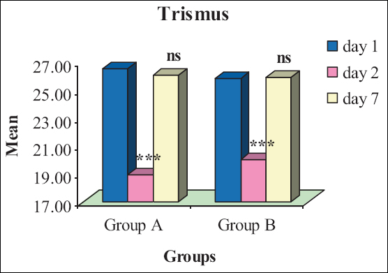
Figure 1: For each group, comparative trismus levels within the groups (i.e., between periods). nsP > 0.0.05 or *** P < 0.001 as compared to day 1


Similarly, for each day, comparing the mean trismus levels between the groups [Table 7], the trismus levels at all periods did not differ (P > 0.05) between the two groups that is, found to be statistically the same.

Table 7: For each period, the significance (P value) of the mean difference of trismus levels between the groups by Tukey test

Periods Comparisons (Group A vs. Group B)
Day 1 0.934
Day 2 0.667
Day 7 1.000
 

Pain

The pain levels before and after treatments for the two groups are summarized in Tables 8 and 9. Both groups experienced an increase in mean pain level after treatment (day 2), followed by a decrease after 7 days, almost returning to the initial levels (day 1).The change was more significant in Group B compared to Group A.

Table 8: Pre and postpain levels (Mean ± SD) of two groups over the periods

Groups Day 1 Day 2 Day 7
Group A 0.35 ± 0.49 (0–1) 4.60 ± 0.88 (4–6) 0.45 ± 0.51 (0–1)
Group B 0.40 ± 0.50 (0–1) 6.60 ± 0.75 (6–8) 0.60 ± 0.50 (0–1)

Numbers in parenthesis indicate the range (min–max)


Table 9: For each period, the significance (P value) of the mean difference of pain levels between the groups by Tukey test

Periods Comparisons (Group A vs. Group B)
Day 1 1.000
Day 2 P < 0.001
Day 7 0.074
 

For each group, comparing the mean pain levels within the groups (i.e., between periods) [Tables 10 and 11], the pain levels in both Groups A and B increased significantly (P < 0.001) at day 2 compared with day 1, whereas decreased significantly (P < 0.001) at day 7 compared with day 2. However, in both groups, the mean pain levels did not differ(P > 0.05) between day 1 and day 7 that is, found to be statistically the same [Table 9 and Figure 2].

Table 10: For each group, the significance (P value) of the mean difference of pain levels with the groups (i.e., between periods) by Tukey test

Comparisons Group A Group B
Day 1 vs. day 2 P < 0.001 P < 0.001
Day 1 vs. day 7 0.996 0.919
Day 2 vs. day 7 P < 0.001 P < 0.001
 

Table 11: Operating time levels (mean ± SD) of two groups

Group A (n = 20) Group B (n = 20) t value (DF=38) P value
47.75 ± 9.81 (32–70) 29.40 ± 4.89 (20–38) 7.49 P < 0.001

***P < 0.001—Group A vs. Group B

 
thumb

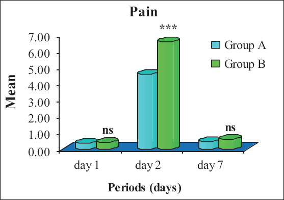
Figure 2: For each day, comparative pain levels between the groups


Operating time

The operating times of the two groups are summarized in Table 11 and also shown graphically in Figure 3. The operating time of Groups A and B ranged from 32 to 70 min and from 20 to 38 min, respectively, with a mean ± SD of 47.75 ± 9.81 and 29.40 ± 4.89 min, respectively. The mean operating time of Group B was comparatively lower than Group A. Comparing the mean operating time of the two groups, t test revealed significantly (P < 0.001) and 38.4% lower operating time of Group B compared with Group A (47.75 ± 9.81 vs. 29.40 ± 4.89, t = 7.49; P < 0.001).

thumb

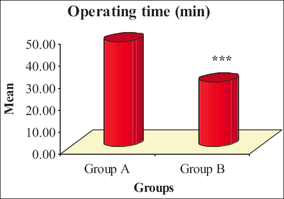
Figure 3: Mean operating time of two groups


DISCUSSION

The study was conducted at the Department of Oral and Maxillofacial Surgery, Teerthanker Mahaveer Dental College and Research Center in Moradabad. Each group consisted of a total of 20 patients who were treated using either the conventional bur or lingual split technique. We evaluated two commonly used procedures for removing impacted mandibular third molars to understand their effectiveness. The study aimed to assess the clinical impact of two different bone-cutting techniques during the extraction of impacted mandibular third molars. Only asymptomatic patients were included and randomly assigned to either the conventional bur technique or the lingual split technique.

Swelling

In previous studies, various methods have been used to assess postoperative swelling. In our current study, we utilized the measurement method from the ear to the corner of the mouth as described by Yuasa and Sugiura.[12] Our findings showed significant swelling on the second day, consistent with observations by Susarla et al.[13] and Shugars et al.[14] Furthermore, it was noted that swelling peaked on the second day for 46% of the patients, began to diminish by the fourth day, and was even less by the seventh day.[15] In our study, Group B exhibited higher swelling compared with Group A on the second day. This increase in swelling for Group B may be attributed to the use of electrically driven instruments, which can push bone particles laden with microorganisms deeper into the bone, potentially causing infection. Additionally, incomplete sterilization of instruments can lead to cross-infection. Furthermore, the use of rotary instruments can generate frictional heat during bone tissue removal, which may impede the healing process.

Trismus and operating time

According to Stanley,[16] needle piercing alone does not cause trismus. However, if the needle accidentally damages the periosteum, it can lead to muscle spasms and trismus. White et al.[17] explain that surgery triggers an inflammatory process, leading to muscle contraction and trismus. In our study, the trismus score was significantly higher in Group A than in Group B. We found that mouth opening was much better in the lower age groups on day 2 and day 5. Additionally, the duration of oral surgery was identified as a predictive factor for pain, trismus, and edema. According to Alkadi and Stassen,[18] the longer the operative time, the higher the likelihood of postoperative complications.

Pain

Postoperative pain develops due to localized inflammation in surgical areas conjugated tissue injury and cellular destruction provoke releasing and production of several biochemical mediators, for example, histamine, bradykinin, and prostaglandins, which are involved in the pain process[19] and the level of postoperative pain decreases with time, reaching its peak on the second and third days after surgery and decreasing to a minimum on the seventh day during suture removal. Our study indicates that Group A experienced less pain compared with Group B. Severe pain was linked to the depth of the tooth and the preoperative index of difficulty. Additionally, facial swelling appeared to be influenced by individual characteristics such as age and sex, as well as by the relationship between the trismus due to the close vicinity of the ramus and the angle of the mandible. Sortino and Cicciù,[20] the enlargement of the incision and manipulation of tissue has the potential to impact the magnitude of swelling and mouth opening. Interestingly, severe swelling was observed in cases of easier extractions, which were associated with a wider relation to the ramus and available space, possibly impacting the patient’s facial shape. Both severe and moderate pain were linked to the depth of the teeth and the difficulty of extraction.

According to Ten Bosch and Van Gool[21] and Van Gool et al.,[22] discomfort peaked within 2–3 days, concurrent with swelling, leading to trismus. By the seventh day, a noticeable improvement in mouth opening occurred. Chiapasco et al.[23] stressed the significance of excessive bone guttering in worsening surgical discomfort. In this study, it was found that techniques using a chisel, specifically the lingual split bone technique were associated with less external swelling than the surgical bur technique. These findings are consistent with the results reported by other authors. On the seventh postoperative day, there was no significant difference in pain between the surgical bur technique and the lingual split bone technique; however, the pain was more pronounced with the lingual split bone technique in comparison to the conventional bur technique.

CONCLUSION

The results of this study are promising, but more research is needed to confirm them and identify risk factors. Ongoing research includes various surgical techniques (cryotherapy, ozone gel, platelet-rich plasma, ice application, cyanoacrylate, platelet-rich fibrin, piezoelectric surgery, and laser surgeries) and genetic studies (involvement of MSX1 and AXIN2 genes). The lingual split technique was found to be more painful, and the surgical bur technique resulted in more swelling. There is still no consensus on the best protocol for extracting impacted lower third molars, often depending on the surgeon’s experience.

Limitations of the study

The study’s limitations include uncontrolled confounding factors such as oral hygiene, age, gender, and systemic health conditions. Additionally, variations in extraction techniques among different dental practitioners were not considered. Furthermore, longitudinal studies in diverse populations are needed to better understand the risk factors for postoperative complications. Surgeons need guidance in decision-making regarding impacted tooth extraction to reduce postoperative problems.

Acknowledgements

I would like to express my heartfelt gratitude for the efforts and dedication put into this research, which was edited and mentored by renowned academician Prof. (Dr.) Arun Goyal Sir, as well as Dr. Uday Dangi Sir, Dr. D. S. Gupta Sir, and Dr. Ravi Jain Sir in the field of Oral and Maxillofacial Surgery.

Data availability statement

The current study’s datasets are available upon reasonable request from the corresponding author.

Ethics committee approval

The Institutional Ethics Committee Teerthankar Mahaveer University Moradabad, meeting held on December 6, 2010, has approved and provided its consent in this regard. The study complies with the STROBE checklist guidelines.

Informed consent

Following the Helsinki Declaration, informed consent was obtained from every participant in this study.

Financial support and sponsorship

Nil.

Conflicts of interest

There are no conflicts of interest.

REFERENCES

1. Saputri RI, De Tobel J, Vranckx M, Ockerman A, Van Vlierberghe M, Fieuws S, et al. Is third molar development affected by third molar impaction or impaction-related parameters?Clin Oral Investig 2021;25:6681-93. [CrossRef] [PubMed] [Google Scholar]

2. Carter K, Worthington S. Predictors of third molar impaction:A systematic review and meta-analysis. J Dent Res 2016;95:267-76. [CrossRef] [PubMed] [Google Scholar]

3. Caillet J, Pereira B, Courtot R, Barthelemy I, Devoize L, Depeyre A. Ectopic third mandibular molar:Evaluation of surgical practices and meta-analysis. Clin Oral Investig 2021;25:4781-99. [CrossRef] [PubMed] [Google Scholar]

4. Zhang Y, Chen X, Zhou Z, Hao Y, Li H, Cheng Y, et al. Effects of impacted lower third molar extraction on periodontal tissue of the adjacent second molar. Ther Clin Risk Manag 2021;17:235-47. [CrossRef] [PubMed] [PubMed Central] [Google Scholar]

5. Aniko-Wlodarczyk M, Jaron A, Preuss O, Grzywacz A, Trybek G. Evaluation of the effect of surgical extraction of an impacted mandibular third molar on the periodontal status of the second molar-prospective study. J Clin Med 2021;10:2655. [CrossRef] [PubMed] [PubMed Central] [Google Scholar]

6. Poszytek D, Gorski B. Relationship between the status of third molars and the occurrence of dental and periodontal lesions in adjacent second molars in the Polish population:A radiological retrospective observational study. J Clin Med 2024;13:20. [CrossRef] [PubMed] [PubMed Central] [Google Scholar]

7. Menager L, Ruperto M, Fricain CJ, Catros S, Fenelon M. Does surgical removal of mandibular third molar influence the periodontal status of the adjacent second molars?A systematic review. J Oral Med Oral Surg 2023;29:1. https://doi.org/10.1051/mbcb/2022032 [Google Scholar]

8. Winter GB. Impacted Mandibular Third Molars. St. Louis, MO:American Medical Book Co.;1926. [Google Scholar]

9. Tetsch P, Wagner W. Operative Extraction of Wisdom Teeth. London:Wolfe Medical Publication;1985. [Google Scholar]

10. Pell GJ, Gregory GT. Impacted mandibular third molars:Classification and modified technique for removal. Dent Dig 1933;39:330-8. [Google Scholar]

11. Asanami S, Kasazaki Y. Expert Third Molar Extractions. Tokyo, Japan:Quintessence Publishing;1990. [Google Scholar]

12. Yuasa H, Sugiura M. Clinical postoperative findings after removal of impacted mandibular third molars:Prediction of postoperative facial swelling and pain based on preoperative variables. Br J Oral Maxillofac Surg 2004;42:209-14. [CrossRef] [PubMed] [Google Scholar]

13. Susarla SM, Blaeser BF, Magalnick D. Third molar surgery and associated complications. Oral Maxillofac Surg Clin North Am 2003;15:177-86. [CrossRef] [PubMed] [Google Scholar]

14. Shugars DA, Benson K, White RP, Jr, Simpson KN, Bader JD. Developing a measure of patient perceptions of short-term outcomes of third molar surgery. J Oral Maxillofac Surg 1996;54:1402-8. [CrossRef] [PubMed] [Google Scholar]

15. Karpe T, Sanober A, Nasyam FA, Soumya S, Seethamsetty S, Sarepally G. To study the impact of tooth sectioning on postoperative pain, swelling and trismus after surgical extraction of impacted mandibular third molars. Cureus 2023;15:e51207. [PubMed] [PubMed Central] [Google Scholar]

16. Stanley MF. Hand book of Local Anaesthesia. 5th ed. St. Louis, MO:Mosby;2004. 308. [PubMed Central] [Google Scholar]

17. White RP, Jr, Shugars DA, Shafer DM, Laskin DM, Buckley MJ, Phillips C. Recovery after third molar surgery:Clinical and health- related quality of life outcomes. J Oral Maxillofac Surg 2003;61:535-44. [CrossRef] [PubMed] [Google Scholar]

18. Alkadi S, Stassen L. Effect of one-suture and sutureless techniques on postoperative healing after third molar surgery. J Oral Maxillofac Surg 2019;77:703.e1-703.e16. [CrossRef] [PubMed] [PubMed Central] [Google Scholar]

19. Kumar BS, Sarumathi T, Veerabahu M, Raman U. To compare standard incision and comma shaped incision and its influence on post-operative complications in surgical removal of impacted third molars. J Clin Diagn Res 2013;7:1514-8. [Google Scholar]

20. Sortino F, Cicciu M. Strategies used to inhibit postoperative swelling following removal of impacted lower third molar. Dent Res J (Isfahan) 2011;8:162-71. [CrossRef] [PubMed] [PubMed Central] [Google Scholar]

21. Ten Bosch JJ, van Gool AV. The interrelation of postoperative complaints after removal of the mandibular third molar. Int J Oral Surg 1977;6:22-8. [CrossRef] [PubMed] [Google Scholar]

22. van Gool AV, Ten Bosch JJ, Boering G. Clinical consequences of complaints and complications after removal of the mandibular third molar. Int J Oral Surg 1977;6:29-37. [CrossRef] [PubMed] [Google Scholar]

23. Chiapasco M, De Cicco L, Marrone G. Side effects and complications associated with third molar surgery. Oral Surg Oral Med Oral Pathol 1993;76:412-20. [CrossRef] [PubMed] [Google Scholar]