Department of Dentistry, ESIC Medical College and PGIMSR, KK Nagar, Chennai, Tamil Nadu, India
Submitted: 02-Sep-2021,
Accepted in Revised Form: 24-Oct-2021,
Published: 29-Dec-2021
DOI: 10.4103/sidj.sidj_15_21
Saint Int Dent J 2021;5:64-7.
Copyright: © 2021 The Saint's International Dental Journal
Licence
This is an open access journal, and articles are distributed under the terms of the Creative Commons Attribution-NonCommercial-ShareAlike 4.0 License, which allows others to remix, tweak, and build upon the work non-commercially, as long as appropriate credit is given and the new creations are licensed under the identical terms.
Disclaimer:
This article was originally published by Wolters Kluwer Medknow Publications & Media Pvt Ltd and has now been officially transferred to Society of Dental Research & Education due to a change in publishing entity.
Oral mucosal trauma can cause a multitude of oral mucosal lesions. Many oral mucosal lesions are caused by prolonged mechanical trauma to the oral mucosa. This case series depicts five such cases in which trauma was the major etiological factor in the causation of such lesions. This case series discusses the five different entities: Traumatic ulcer, frictional keratosis, traumatic fibroma, mucocele, and angina bullosa hemorrhagica. The etiology, clinical features, and management of each of these lesions are discussed. Appropriate symptomatic management of mucosal lesions along with the removal of the etiological factor causing trauma to the oral cavity resulting in the complete resolution of all five oral lesions described in this case series. This case series aims to illustrate the need for early diagnosis of trauma-associated oral mucosal lesions to avoid broad therapy later on.
Keywords: Mucosa, oral lesion, trauma, ulcer
Oral mucosal trauma characterized by a defect in underlying connective tissue, epithelium, or both has been a growing concern in the recent decade as trauma may result in consequences ranging from a small oral ulcer to a malignant lesion. A variety of oral mucosal disorders induced by acute and chronic trauma can be visible in the oral mucosa, including acute or chronic ulcers, white or red lesions, mucositis, reactive hyperplasia, and even oral ulcerations with bone sequestration.[1] Physical, chemical, or thermal trauma can lead to injury of the oral mucosa.[2] Chronic mechanical irritation to the oral mucosa is produced through low-intensity, sustained, and repeated action of an oral deleterious agent, which teeth, dentures can cause, and functional alterations, either through separate, or combined action. Prompt diagnosis and elimination of the traumatic etiological feature are key to managing such trauma-associated oral mucosal lesions. This case series enumerates some oral mucosal abnormalities reported to the Department of Dentistry, where trauma was considered the major etiological factor.
A 58-year-old female reported with a chief complaint of frequent oral wounds on the right side of the tongue for 4 months. Presently, she complained of a painful oral wound on the right side of the tongue for 1 week. On examination, two predominant oral ulcers measuring 0.7 cm and 0.4 cm approximately were present in the tongue’s right lateral border in relation to #46 region [Figure 1a]. Two more healing ulcers were also present distal to the present ulcers connoting the frequent oral ulcerations. On palpation, the ulcers were tender and nonindurated in nature. No bleeding on palpation was present. Hard-tissue examination revealed a grossly carious root of #46, traumatizing the tongue. Based on the history and clinical findings, the patient was provisionally diagnosed with traumatic ulcers in the right lateral border of the tongue. The patient was prescribed topical analgesics and anti-inflammatory agents for symptomatic relief of pain caused by oral ulcers. Extraction of 46 was done under local anesthesia. The patient was reviewed after 2 weeks; there was complete healing of traumatic oral ulcers [Figure 1b].
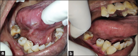
Figure 1: Traumatic ulcer of right lateral border of tongue – (a) Pretreatment. (b) Posttreatment
A 49-year-old male presented with the chief complaint of a sharp tooth impinging the tongue in the left side of the tongue. On examination, there was a white keratotic lesion of 1 cm × 0.5 cm present in the left lateral border of the tongue [Figure 2]. The hard-tissue examination revealed an attrited sharp cusp in relation to #36 which was traumatizing the left lateral border of the tongue. Based on the history and clinical findings, a provisional diagnosis of frictional keratosis was arrived. The occlusal grinding of the sharp cusp was done and the patient is under periodic follow-up.
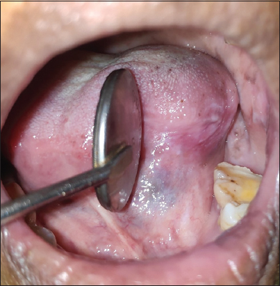
Figure 2: Frictional keratosis of left lateral border of tongue
A 28-year-old female complained primarily of pain and swelling in the inner part of the left cheek region, noted over 3 weeks. In the beginning, the pain was gradual and worsened while eating. On examination, there was a solitary, well-defined sessile growth of 1.0 cm × 0.5 cm with surface ulceration present in the posterior buccal mucosa in relation to the #28 region [Figure 3a]. The growth was tender, firm inconsistency, and not indurated on palpation. Hard-tissue examination revealed a buccoverted, grossly decayed 28 impinging, and traumatizing the growth [Figure 3b]. Based on the history and clinical findings, the patient was provisionally diagnosed with traumatic fibroma of the left buccal mucosa in relation to the #28 region. The preliminary examination was performed, and #28 was extracted, followed by an excisional biopsy of the lesion. Histopathological examination of soft tissue revealed dense fibrous tissue in the subepithelium with capillaries.
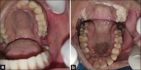
Figure 3: (a) Traumatic fibroma ofleftbuccal mucosa. (b) Buccoverted grossly decayed 28 traumatizing the left buccal mucosa visualized in palatal mirror
A 23-year-old male presented with a chief complaint of swelling in the inner side of the lower lip for 1 month. The patient gave a history of accidentally biting the lip while having food before the onset of swelling. The swelling was not painful, and it was initially small, and it has progressed to the present size. On examination, there was a solitary diffuse swelling of approximately 1.0 cm × 1.5 cm was seen in the lower labial mucosa corresponding to #32, #33 region [Figure 4]. The swelling was nontender and soft inconsistency. A provisional diagnosis of mucocele of the lower labial mucosa was made based on the history and clinical findings. Following basic tests, an excisional biopsy of the lesion was conducted under local anesthetic, and the histological findings were congruent with mucocele.
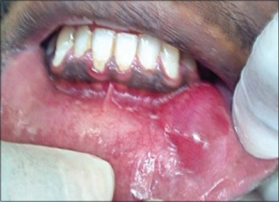
Figure 4: Mucocele of lower labial mucosa
A 45-year-old female had the main complaint of a sudden reddish swelling on the left side of the tongue from a day earlier. The patient gave a history of hard food intake before the onset of the lesion. The patient had no bleeding disorders in the past, and there was no history of recurrence of such swelling. On examination, a red-colored blood-filled blister of about 1 cm in diameter was present in the left lateral border of the tongue [Figure 5]. On palpation, the lesion was sessile, nontender, and soft in consistency. The provisional diagnosis of angina bullosa hemorrhagica (ABH) was made based on the clinical appearance and history of the reddish swelling on the tongue’s left lateral border. Laboratory evaluation of complete blood count and coagulation profile did not reveal any underlying disease and gave a normal picture. The healing was uneventful as it ruptured spontaneously within a day. Topical oral steroids and anti-inflammatory agents were prescribed for symptomatic relief.
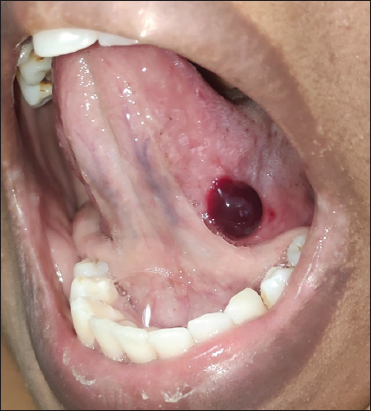
Figure 5: Angina bullosa hemorrhagica of left lateral border of tongue
In this case series, five patients with trauma-induced oral mucosal pathologies were included, out of which three were female and two were male. The age of the patients in the present case series ranged from 23 to 58 years. In all the patients, trauma was the major etiological factor that resulted in the oral mucosal pathologies [Table 1]. Removing the etiological factor that caused trauma in the oral cavity followed by prompt treatment resulted in the complete resolution of these oral pathologies in all the cases presented in this case series. This discussion would enumerate the five different trauma-associated oral mucosal lesions described in this case series. Trauma-induced oral mucosal lesions may be symptomatic or asymptomatic. Asymptomatic oral mucosal lesions are sometimes diagnosed at a later stage or accidentally diagnosed during an intraoral examination.
Table 1: Demographic data, clinical details, and management of five patients with trauma-induced oral mucosal pathologies
| Patient | Age | Gender | Etiology | Diagnosis | Treatment |
|---|---|---|---|---|---|
| 1 | 58 | Female | Trauma due to grossly decayed carious root of #46 | Traumatic ulcers of right lateral border of tongue | Symptomatic management of the ulcers and extraction of #46 |
| 2 | 49 | Male | Trauma due to attrited #36 | Frictional keratosis of left lateral border of tongue | Occlusal grinding of the sharp cusp of #36 |
| 3 | 28 | Female | Trauma due to grossly decayed buccoverted #28 | Traumatic fibroma of left buccal mucosa | Extraction of #28 followed by excisional biopsy of the traumatic fibroma |
| 4 | 23 | Male | Trauma due to lip biting | Mucocele of lower labial mucosa | Excisional biopsy of the mucocele |
| 5 | 45 | Female | Trauma due to hard foods | Angina bullosa hemorrhagica of left lateral border of the tongue | Spontaneous rupture |
The most common lesions that are caused by chronic mechanical irritation are tongue/cheek biting, frictional keratosis, indentations, chronic traumatic ulcer, papillary hyperplasia, denture-induced fibrous hyperplasia, and focal fibrous hyperplasia.[3] Traumatic ulcers usually present as solitary lesions that have a nonspecific clinical shape. Traumatic ulcer can also be classified as a chronic solitary ulcer because chronic injuries of oral mucosa usually predispose to such ulcers.[4] Frictional keratosis is seen as a white, keratotic lesion due to chronic mechanical irritation caused by sharp edges of teeth or restorations, dental prosthesis, abrasive foods, vigorous tooth brushing, and playing wind instruments.[5]
A fibroma results from a chronic repair process that includes granulation tissue and scar formation resulting in a fibrous submucosal mass.[6] The common sites of traumatic fibroma are the tongue, buccal mucosa, and lower labial mucosa. Fibromas are broad-based lesions and are relatively lighter in color than the surrounding normal tissue. The surface often appears white because of hyperkeratosis or with surface ulceration caused by secondary trauma.[7] They are usually benign and asymptomatic lesions. The initial treatment involves the identification of the causative local irritative factor followed by their elimination with subsequent monitoring and finally surgical excision.[8] Oral Mucoceles are frequently seen affecting young people, and the lower lip is the most common site for the occurrence of mucoceles. It usually measures around 5–14 mm, and the extravasation type of mucocele is the most common type.[9] Lower lip is the most common site of these lesions in the oral cavity, and the trauma or habit of lip biting is the most postulated cause.[10] If left without intervention, an episodic decrease and increase in size may be observed, based on rupture and subsequent mucin production.[11] Conventional treatment is surgical extirpation of the surrounding mucosa and glandular tissue down to the muscle layer.[12]
Angina Bullosa Hemorrhagica (ABH) is the term used to describe benign subepithelial oral mucosal blisters filled with blood.[13] Badham described ABH to describe oral blood-filled vesicles or bullae that could not be attributed to a blood dyscrasia vesiculobullous disorder, systemic disease, or other known causes.[14] These patients present with a history of oral blood blisters, which occurs following mild oral trauma. The subepithelial blisters rupture, typically during meals, releasing fresh blood, leaving erosion. No involvement of other mucosal membranes or organs is seen.[15] During the workup, laboratory testing, including differential blood count and coagulation assessment tests, are indicated to exclude any possible underlying hemostatic defects. A tissue biopsy or analysis of blister fluid is not advised, as it may cause a secondary infection at the blister site.[16] In the event of the persistence of traumatic ulcers for more than 2 weeks, even after elimination of the traumatic etiological agent that caused it, a biopsy of the lesion is essential to rule out any malignant changes associated with the lesion.[17]
Complete periodic oral examinations are essential because early detection of asymptomatic oral lesions reduces the morbidity associated with such lesions. Replacement of missing teeth to avoid supraeruption of teeth and early treatment of carious tooth is essential to prevent traumatic injuries in the long run. An early diagnosis followed by prompt care is of utmost essential to address the trauma associated with oral lesions. Moreover, in-depth knowledge about oral lesions is essential for the proper diagnosis and prompt care. If the etiological sources of trauma are not eradicated, the risk of recurrence of such lesions cannot be ruled out.
The authors certify that they have obtained all appropriate patient consent forms. In the form the patient(s) has/have given his/her/their consent for his/her/their images and other clinical information to be reported in the journal. The patients understand that their names and initials will not be published and due efforts will be made to conceal their identity, but anonymity cannot be guaranteed.
Nil.
There are no conflicts of interest.
1. Anura A. Traumatic oral mucosal lesions:A mini review and clinical update. Oral Health Dent Manag 2014;13:254-9. [CrossRef] [Google Scholar] [PubMed]
2. Phore S, Panchal RS. Traumatic oral lesions:Pictorial essay. Med J DY Patil Vidyapeeth 2018;11:94. [CrossRef] [Google Scholar]
3. LazosJP, Piemonte ED, Lanfranchi HE, Brunotto MN. Characterization of chronic mechanical irritation in oral cancer. Int J Dent 2017;2017:6784526. [CrossRef] [Google Scholar] [PubMed Central] [PubMed]
4. Mortazavi H, Safi Y, Baharvand M, Rahmani S. Diagnostic features of common oral ulcerative lesions:An updated decision tree. Int J Dent 2016;2016:7278925. [CrossRef] [Google Scholar] [PubMed Central] [PubMed]
5. van der Wal JE. Frictional keratosis. In:van Krieken J, editor. Encyclopedia of Pathology. Cham:Springer;2017. [CrossRef] [Google Scholar]
6. Esmeili T, Lozada-Nur F, Epstein J. Common benign oral soft tissue masses. Dent Clin North Am 2005;49:223-40. [CrossRef] [Google Scholar] [PubMed]
7. Rathva VJ. Traumatic fibroma of tongue. BMJ Case Rep 2013;2013:bcr2012008220. [CrossRef] [Google Scholar] [PubMed Central] [PubMed]
8. Guerrieri P, Oliveira A, Arosio F, Murgia MS, ViganòL, Casu C. Exophytic soft tissue traumatic lesions in dentistry:Asystematic review. Int J Appl Dent Sci 2020;6:387-92. [CrossRef] [Google Scholar]
9. More CB, Bhavsar K, Varma S, Tailor M. Oral mucocele:Aclinical and histopathological study. J Oral Maxillofac Pathol 2014;18:S72-7. [CrossRef] [Google Scholar]
10. Chaitanya P, Praveen D, Reddy M. Mucocele on lower lip:Acase series. Indian Dermatol Online J 2017;8:205-7. [CrossRef] [Google Scholar]
11. Nallasivam KU, Sudha BR. Oral mucocele:Review of literature and a case report. J Pharm Bioallied Sci 2015;7:S731-3. [CrossRef] [Google Scholar] [PubMed Central] [PubMed]
12. Ata-AliJ, Carrillo C, Bonet C, BalaguerJ, Peñarrocha M, Peñarrocha M. Oral mucocele:Review of the literature. J Clin Exp Dent 2010;2:e18-21. [CrossRef] [Google Scholar]
13. Singh D, Misra N, Agrawal S, Misra P. Angina bullosa haemorrhagica. BMJ Case Rep 2013;2013:bcr2012008505. [CrossRef] [Google Scholar]
14. Giuliani M, Favia GF, Lajolo C, Miani CM. Angina bullosa haemorrhagica:Presentation of eight new cases and a review of the literature. Oral Dis 2002;8:54-8. [CrossRef] [Google Scholar] [PubMed]
15. Grinspan D. Angina bullosa haemorrhagica. Int J Dermatol 1999;38:525-8. [CrossRef] [Google Scholar] [PubMed]
16. Peters JP, van Kempen PM, Robijn SM, Thomeer HG. Angina bullosa hemorrhagica:Post-traumatic swelling in the oral cavity –A case report. J Adv Oral Res 2020;11:97-100. [CrossRef] [Google Scholar]
17. Apriasari ML. The management of chronic traumatic ulcer in oral cavity. Dent J (Majalah Kedokteran Gigi) 2012;45:68-72.[Google Scholar] [CrossRef]