1Department of Oral Medicine and Radiology, Anil Neerukonda Institute of Dental Sciences, Visakhapatnam, Andhra Pradesh, India, 2Department of Oral Medicine and Radiology, Narayana Dental College, Nellore, Andhra Pradesh, India, 3Private Practitioner, Nellore, Andhra Pradesh, India
Address for correspondence: Dr. Gadadasu Swathi, Department of Oral Medicine and Radiology, Anil Neerukonda Institute of Dental Sciences, Visakhapatnam, Andhra Pradesh, India. E-mail: gadadasuswathi5@gmail.com
Submitted: 27-Sep-2020,
Accepted in Revised Form: 05-Dec-2020,
Published: 18-Jun-2021
DOI: 10.4103/sidj.sidj_41_20
Saint Int Dent J 2021;5:28-31
Copyright: © 2021 The Saint's International Dental Journal
Licence
This is an open access journal, and articles are distributed under the terms of the Creative Commons Attribution-NonCommercial-ShareAlike 4.0 License, which allows others to remix, tweak, and build upon the work non-commercially, as long as appropriate credit is given and the new creations are licensed under the identical terms.
Disclaimer:
This article was originally published by Wolters Kluwer Medknow Publications & Media Pvt Ltd and has now been officially transferred to Society of Dental Research & Education due to a change in publishing entity.
Juvenile ossifying fibroma is an uncommon fibro-osseous lesion of jaws nature of short-term rapid growth characterized by excessive growth of bone, destruction, and more probable to reoccur. This lesion has more predominance to occur in younger individuals than older ones and mostly involves facial bones. The lesion mostly involves jaws, orbit, cranial bones, ethmoidal complex, skull base, and Calvaria. Radiographically appears as unilocular or multilocular appearance. The lesion was divided into psammomatoid and trabecular types histopathologically. The psammomatous type of juvenile psammomatoid ossifying fibroma principally involves the bones of the orbit and paranasal sinuses. This lesion is considered to have a higher recurrence rate than its counterparts, so it should be taken care of. This is a case report of a 17-year-old female patient referred to our clinic for a swelling complaint and was provisionally diagnosed as a periapical cyst. The radiograph of this swelling appeared as a multilocular mixed lesion upon viewing. An incisional biopsy was performed to confirm the diagnosis, ensuring it was a juvenile ossifying psammomatoid fibroma. The treatment for the lesion was surgical excision and succeeded by restoration. Lesions with different clinical presentations should be viewed cautiously, taken care of for a proper diagnostic approach, and should be followed for more extended periods to avoid recurrence.
Keywords: Juvenile, margins of excision, ossifying fibroma, panoramic radiography, X-ray film
Juvenile ossifying fibroma (JOF) is a type of fibro-osseous lesion most probably occurring in the skull’s cranial bones, calvaria, ethmoidal complex but can be locally aggressive, rarely occurring in jaws. The most common occurrence is the younger population with equal predominance for males and females.[1] According to their microscopic picture structure and newly released WHO classification 2017, there were three main types in the histopathological picture: Cemento Ossifying fibroma (COF), Juvenile Ossifying Psammomatoid (JPOF), and Juvenile Ossifying Trabecular Fibroma (JTOF).[2]
A 17-year-old female patient approached the department of oral medicine with the chief complaint in the right lower jaw for the past year. She reported a history of sudden onset of swelling, which was initially peanut in size, consequently increased to present magnitude. There was no history of pain, fever associated with swelling. Previous medical, dental, and family history was noncontributory. Extraoral examination revealed asymmetry in the right lower one-third of the face due to diffuse solitary swelling of size 2 cm × 2 cm, roughly oval in shape, extending anteroposteriorly from 2 cm
posterior to symphysis 1 cm anterior to the lower right side angle of the mandible. On palpation, mild paresthesia on the right side mandibular premolar region was noticed. On intraoral examination [Figure 1], a grossly decayed tooth of #46 was detected with a positive note of tenderness on vertical percussion. There was a solitary diffuse irregular swelling in lower right mandible vestibular region which is of size 2 cm × 2 cm extending anteroposteriorly from distal aspect of #44 to mesial aspect of #47. Superiorly from the crest of the alveolar bone to the buccal vestibule inferiorly from #44 to #47. Mucosa over swelling has no color changes. On palpation, there was buccal cortical plate expansion from #44 to #47 and lingual cortical plate expansion in relation to #46. Eggshell crackling was seen on the buccal aspect in relation to 46 region.
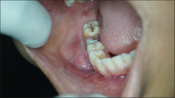
Figure 1. Intraoral swelling
According to clinical findings and observations, provisional of periapical cyst of #46 was given with differential diagnosis of ameloblastoma, dentigerous cyst, calcifying epithelial odontogenic tumor (CEOT) and central ossifying fibroma. The patient was addressed to the radiology department where intraoral periapical (IOPA) radiograph of #46 tooth (IOPA), mandibular right lateral occlusal radiograph, and orthopantomography were taken. IOPA revealed dental caries involving pulp region with loss of distal half of coronal part of #46 was noticed. There was a diffuse radiolucency at periapical region extending from distal half of radicular portion of #45 tooth to distal aspect of #47 tooth running peripherally with sclerotic border, which was visible at the apical region of #45 tooth with evidence of internal septa in #45 and #46 interdental regions.
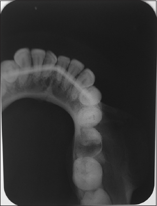
Right lateral mandibular occlusal radiograph depicted buccal cortical plate expansion from #44 to #47 tooth region with the internal structure of mixed radio-opaque and radiolucent structure intermingled with multiple internal septa [Figure 2]. Orthopantamograph revealed solitary well defined, round, multilocular, a mixed radiolucent-radio opaque lesion in right lower mandibular region extending anteroposteriorly from #44 to #47 tooth region apically and superior-inferiorly from the alveolar crest to lower border of the mandible. The mixed
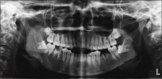
Figure 2. Orthopantamograph
lesion contained many internal septa granting multilocular pattern and displacing the mandibular canal inferiorly. Loss of coronal portion with radicular remnants in relation to #36 was noticed. Radiographic diagnosis of central ossifying fibroma with a differential of multilocular ameloblastoma, CEOT, central hemangioma was recommended [Figure 3].
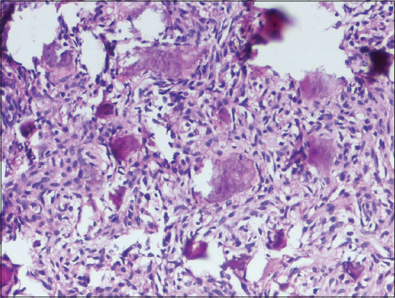
Fine-needle aspiration was performed to find internal content of swelling where the drawing was negative. The patient was subjected to incisional biopsy, the histopathological examination revealed the underlying hypercellular connective tissue of proliferating spindle to fusiform fibroblast arranged in a haphazard manner with discrete deposition of immature bone arranged in a spherical, crescent-shaped like psammomatoid manner and few are in trabecular form with osteoblastic rimming and osteocytes within it, arriving at the diagnosis of psammomatoid juvenile ossifying fibroma [Figure 4]. Bearing in mind, the size and aggressiveness and recurrence rate of the lesion segmental resection was planned and carried out.
Johnson et al. in 1991 postulated that psammamatoid JOF as a cellular mass, generating a varied number of tiny similar osteoid bodies. Overproduction of myxofibrous cellular stroma may be involved in the development of septa in paranasal sinuses during their growth might be the etiology of JOF as suggested by Fletcher et al.[3] JOF most frequently manifests between the age group of 5 and 15 years. JTOF predominates in the age group of 0.5–12 years whereas JPOF preponderant in the age group of 16–33 years. Male population principally the most affected than the female population.[4,5]
JPOF predominates in extragnathic craniofacial bones such as periorbital, ethmoidal, frontal bones, and paranasal sinus bone wall. JTOF prevails mostly in maxilla succeed by mandible though the extragnathic occurrence is sporadic.[6] JOF manifests as an asymptomatic bony swelling. Pain and paraesthesia are a rare phenomenon. In the early stages, the pathognomic finding is the displacement of the adjacent tooth and facial asymmetry and jaw deformity in advanced
Figure 3. Mandibular lateral occlusal radiograph
cases. When JOF has extragnathic presentation, diplopia, exophthalmos, disturbance invisibility, nasal obstruction, and epistaxis can be noticed.[7] Complications mainly arise due to the secondary effect of aggressive growth, thereby encroachment on adjacent anatomical structures, resulting in the associated issues.[8] JOF must be differentiated from other ossifying fibromas that replace the bone with cellular tissue. These include fibrous dysplasia (FD), cement-osseous dysplasia (COD) and osteosarcoma.[9] JOF is mostly a well-demarcated expansile lesion and mostly mixed lesion depending on mineralized structure within the lesion, unilateral in distribution, due to aggressive growth, thinning of cortical plates and consequently, perforation can be noticed. JPOF mostly represents ground-glass appearance, but JTOF depicts radiolucent or mixed lesion. FD, osseous dysplasia, odontoma and ameloblastoma resemble JOF radiographically.[10]
Histologically JOFs represent the proliferation of fibrocellular tissue followed by mineralization. The aggressive nature of growth can be depicted by the presence of intense mitotic figures, sometimes can be mistaken for malignancy. Lack of cellular atypia and the regular mitotic picture depict the benign character of the lesion.[11] JOF histopathologically divided into psammomatoid and trabecular patterns. JPOF characterized by multiple spherical ossicles resembling psammoma bodies, though JTOF is rich in trabeculae of woven bone with coarse lacunae, swollen osteocytes, and a lining of plump osteoblasts. Pathologically COF, FD, COD are a differential diagnosis of JOF. COF occurs in older individuals.[3] It was postulated that JOF can have a recurrence rate of 30%–58% henceforth complete surgical excision should be planned to avoid any recurrences. Malignant transformation has not been reported to date.[12] Clinically, paresthesia is the rare entity in JOF, though present in the present case. Due to its high recurrence rate, after carrying out an incisional
Figure 4. Histopathological picture showing with multiple psammoma bodies
biopsy, segmental excision of the lesion and the patient was followed up to 1 year with no abnormality.[13]
JOF occurring in the mandible is a rare entity, clinical diagnosis was given as a radicular cyst that turned to the multilocular mixed lesion in theradiograph and arose as JOF on histopathological examination. Although JOF is considered to be benign, its aggressive growth turns treatment protocol a bit troublesome. The planned diagnostic approach plays a vital role as JOF’s aggressiveness can be mistaken as a malignant tumor, though it is benign in nature. Clinical management comprises of early diagnosis and long-term follow-up schedules to decrease the severity of the condition.
The authors certify that they have obtained all appropriate patient consent forms. In the form, the patient has given her consent for her images and other clinical information to be reported in the journal. The patient understands that her name and initials will not be published and due efforts will be made to conceal identity, but anonymity cannot be guaranteed.
We would like to acknowledge all patients who gave their acceptance to present their pictures.
Nil.
There are no conflicts of interest.
1. Fatma S, Das SN, Singh S, Rath R. Juvenile trabecular ossifying fibroma of the mandible. Oncol J India 2019;3:16-9. [CrossRef] [Google Scholar]
2. Nguyen S, Hamel MA, Chénard-Roy J, Corriveau MN, Nadeau S. Juvenile psammomatoid ossifying fibroma:A radiolucent lesion to suspect preoperatively. Radiol Case Rep 2019;14:1014-20. [CrossRef] [Google Scholar] [PubMed Central] [PubMed]
3. Fletcher CD. Diagnostic histopathology of tumors. Vol. 1, 3rd ed. Philedelphia, USA:Churchhill Livingstone Elsivier Publication;95-6. [CrossRef] [Google Scholar]
4. Nair P, Kumar A, Hegde K, Neelakantan S. Psammomatoid juvenile ossifying fibroma:A case report with literature review. J Indian Acad Oral Med Radiol 2010;22:S53-7. [CrossRef] [Google Scholar]
5. Aslan F, Yazici H, Altun E. Psammomatoid variant of juvenile ossifying fibroma. Indian J Pathol Microbiol 2018;61:443-5. [CrossRef] [Google Scholar] [PubMed]
6. SultanAS, Schwartz MK, Caccamese JF Jr., Papadimitriou JC, Basile J, Foss RD, et al. Juvenile trabecular ossifying fibroma. Head Neck Pathol 2018;12:567-71. [CrossRef] [Google Scholar]
7. El-Mofty SK, Nelson B, Toyosawa S. Ossifying fibroma. In:El-Naggar AK, Chan JK, Grandis JR, Takata T, Slootweg PJ, editors. WHO Classification of Head and Neck Tumours. 4th ed.. Lyon:IARC Press;2017. 251-2. [CrossRef] [Google Scholar]
8. Malaviya P, Choudhary S, Gupta S, Toshniwal OD. Trabecular variant:A rare entity of juvenile ossifying fibroma of the mandible. Contemp Clin Dent 2017;8:179-81. [CrossRef] [Google Scholar] [PubMed Central] [PubMed]
9. Kwon Y, Shin D, Kim J, Lee M, Choi H. Juvenile psammomatoid ossifying fibroma of the maxilla. Arch Craniofac Surg 2020;21:193-7. [CrossRef] [Google Scholar] [PubMed Central] [PubMed]
10. Lemoine S, Cassagnau E, Bertin H, Poisson M, Corre P, GuiolJ. Juvenile ossifying fibroma:Case report and literature review, management and differential diagnosis. J Oral Med Oral Surg 2018;24:67-71. [CrossRef] [Google Scholar]
11. Han J, Hu L, Zhang C, Yang X, Tian Z, Wang Y, et al. Juvenile ossifying fibroma of the jaw:A retrospective study of 15 cases. Int J Oral Maxillofac Surg 2016;45:368-76. [CrossRef] [Google Scholar]
12. Diniz JA, Siqueira AD, Araujo GM, Faro TF, Torres LH, Oliveira E SilvaED, et al. Intraoral approach for surgical treatment of psammomatoid juvenile ossifying fibroma. J Craniofac Surg 2020;31:e306-9. [CrossRef] [Google Scholar] [PubMed]
13. Martinez-Manas RM, Rey MJ, Gaston F. Aggressive psammomatoid ossifying fibroma. Ann Otol Rhinol Laryngol 2002;111:466-8. [CrossRef] [Google Scholar] [PubMed]