1Department of Periodontics, Sathyabama Dental College and Hospital, Chennai, Tamil Nadu, India, 2Department of Conservative Dentistry and Endodontics, CSI College of Dental Sciences and Research, Madurai, Tamil Nadu, India
Address for correspondence: Dr. Yamini Rajachandrasekaran, Department of Periodontics, Sathyabama Dental College and Hospital, Chennai, Tamil Nadu, India. E-mail: yammu.cool@gmail.com
Submitted: 12-Apr-2020
Revised: 30-Apr-2020
Accepted: 08-May-2020
Published: 28-Jul-2020
DOI: 10.4103/sidj.sidj_12_20
Saint Int Dent J 2020;4:67-70.
Copyright: © 2020 The Saint's International Dental Journal
Licence
This is an open access journal, and articles are distributed under the terms of the Creative Commons Attribution-NonCommercial-ShareAlike 4.0 License, which allows others to remix, tweak, and build upon the work non-commercially, as long as appropriate credit is given and the new creations are licensed under the identical terms.
Disclaimer:
This article was originally published by Wolters Kluwer Medknow Publications & Media Pvt Ltd and has now been officially transferred to Society of Dental Research & Education due to a change in publishing entity.
Planning a course of treatment for periodontally compromised patients is a multidisciplinary approach. The goal of treatment should aim at achieving the physiologic form, esthetics, and function of all teeth and also to stabilize the tooth mobility by splinting if the teeth are to be preserved. Hence, replacing a missing tooth in a periodontally compromised patient involves the joint collaboration of periodontist, prosthodontist, and endodontist, and, at times, orthodontists are involved to achieve the treatment goal. This case presents management of missing tooth in periodontally compromised patients (periodontal prosthetic ptients) using indirect fiber-reinforced prosthesis which acts as periodontal prosthesis.
Keywords: Fiber-reinforced composite bridge, periodontally compromised teeth, ridge augmentation, splints, tooth mobility
Localized alveolar ridge defect refers to volumetric deficit of the bone and soft tissue within the alveolar process, and it is frequently found in partially edentulous patients. The alveolar process often undergoes a continuous resorptive process and seems to follow a predictable pattern where it first reduces in width and later in height.[1,2] There is a high incidence of residual ridge deformity following anterior tooth loss.[3] Alveolar ridge deformity occurs due to traumatic teeth removal, severe periodontal diseases, endodontic failure, implant failure, traumatic accidents, and developmental defects.
Siebert classified residual ridge deformity into the following three categories: Class I is characterized by the faciolingual loss of the tissue with normal ridge height. Class II involves the loss of ridge height with normal ridge width. Class III is marked by a combination of loss in both dimensions. Class I is not esthetically challenging. However, Class II and Class III present more difficulty as it involves in the reconstruction of a high volume of tissue. Various grafting procedures have been developed such as soft-tissue autografts[4,5] and hard-tissue grafts.[6]
This case report describes a multidisciplinary approach to a prostheic treatment in periodontally compramised patients where ridge augmentation is followed by periodontal-supported prosthesis.
A 37-year-old woman reported to the department with a chief complaint of replacing her missing teeth in the lower front tooth region of the jaw. Her past dental history revealed that she had undergone extraction of her mobile teeth before 1 year. The patient is a known diabetic for the past 2 years and under control. A detailed family history was obtained, and it was unremarkable.
Clinical examination revealed the presence of all teeth except the lower left mandibular incisor. Miller’s[7] Grade II mobility with a clinical attachment level of 6 was evident in the right lower central incisor. On probing, there was a generalized pocket depth of about 4–7 mm, and she had a score of fair on oral hygiene examination.[8] On examination of the gingiva, there was generalized roundening of the marginal gingiva and ballooning of interdental papilla was evident. Radiographic examination revealed generalized horizontal bone loss.
The treatment plan was aimed at improving the patient’s overall periodontal health as well as to restore the lost tooth. A thorough clinical and radiographical examination revealed Siebert[9] Class III ridge deformity [Figure 1] and further examination revealed that the patient’s palate had sufficient thickness to serve as a donor site.
Figure 1: Preoperative photographs
Hence, ridge augmentation was planned for periodontal prosthesis as the fixed prosthesis is not feasible for the patient due to periodontally compromised teeth. Furthermore, the patient was not opting for the extraction of mobile teeth as well as the implant. Free gingival graft (FGG) itself fulfills the necessary requirements for periodontal prosthesis.
Hence, considering the technique sensitivity for connective tissue graft procedure compared to FGG, FGG was preferred. FGG along with hard-tissue substitutes such as xenografts was planned for ridge augmentation in the edentulous site.
Phase I therapy comprising of scaling, polishing, subgingival root planing, and correction of occlusal disharmony had been followed and revaluated for 4 weeks. In the surgical phase, convention flap surgery was carried out in all the four quadrants in stages, followed by which the soft- and hard-tissue augmentation was carried out.
After administering 2% lignocaine (1: 80,000) (Lignox 2% A Indoco, India), horizontal incisions were given on the lingual aspect of edentulous site 2 mm apical from the crest of the ridge [Figure 2]. Horizontal incision is joint with the sulcular incision extending to 32 and 41, and a full-thickness flap was elevated [Figure 2]. A FGG measuring about 8 mm × 7 mm × 6 mm was obtained from the upper right palatal region. Then, the xenograft (Advanced Biotech Products [P] Ltd.,) particles were placed, followed by the FGG acting as a membrane in the recipient site and secured by suturing (Johnson and Johnson Pvt. Ltd., India) with the flap, and the flap was approximated with interrupted sutures (Johnson and Johnson Pvt Ltd., India) [Figure 3].
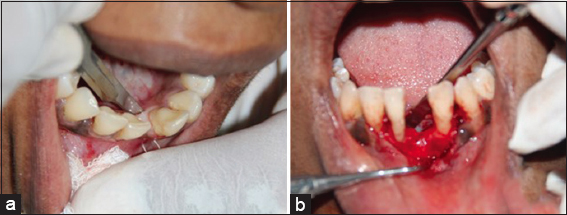
Figure 2: Incision placement and flap retraction
Figure 3: Graft placement and suture placement
Follow-up was done after 1 week, 4 weeks, and 3 months. At the 3rd-month review, a considerable gain in the ridge both apicocoronally and buccolingually was obtained [Figure 4]. After 3-month follow-up, splinting with preimpregnated fiberglass ribbon (EverStick® C and B; GC EUROPE N.V., Leuven, Belgium) along with replacing 31 using flowable composite tooth through indirect method was done where 43, 42, and 33 were used as abutment for stabilizing the periodontally compromised teeth [Figure 5].
Figure 4: Postoperative at 3 months
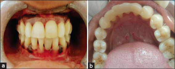
Figure 5: Fiber-reinforced splint along with indirect method of composite tooth placement
Periodontal disease is a complex condition. The incidence of prosthetic treatment in patients with reduced periodontal support is constantly on the rise. Managing a periodontal prosthetic patient always involves a multidisciplinary approach. The goal of the treatment should achieve the physiologic form, esthetic, and function of all the teeth and also to control the tooth mobility of periodontally compromised teeth by splinting.[10]
In the present case, the patient presented with a missing tooth along with periodontally compromised mobile teeth as the adjacent teeth. As the patient was not opting for the extraction as well as for the implant placement, a multidisciplinary approach aiming to replace the missing tooth esthetically with natural emergence profile as well as to preserve the periodontally compromised mobile teeth was planned. Splinting periodontally affected teeth helps in faster healing, and regenerative procedures such as augmentation have greater predictability if the tooth movement is eliminated.[11] In the present case, 43, 32, and 33 were selected as abutment as they served the Ante’slaw[12] for the selection of the abutment for splinting. Conventional fixed partial denture with few modifications or a resin-bonded fixed partial denture could act as a splint along with replacing a missing tooth.[11] Optionally, resin-bonded splints can be designed if the anatomy and situation are not conductive to stand the cement-retained fixed prosthesis. In such situation, fiber-reinforced composite bridges represent an alternative. It can be done directly[13-15] or indirectly. In the direct method, a plastic tooth or avulsed or extracted teeth can be used, and in indirect method, direct tooth built-up is done using composite resin. In our case report, we have employed an indirect method using a preimpregnated fiberglass ribbon (EverStick® C and B; GC EUROPE N.V., Leuven, Belgium).
As the edentulous site presented with localized ridge defect of Siebert Class III defect, to provide an esthetic natural emergence profile for the pontic, soft- and hard-tissue augmentation was performed 3 months prior to the periodontal prosthesis placement. At 6-month follow-up, the restoration was still in place.
Reasonable esthetic appearance was obtained. Fiber re-inforced composite Maryland-like bridge can be considered as a periodontal prosthesis for prosthetic replacement in patients with reduced periodontal support. Furthermore, evaluation needs to be done on a long-term basis.
The authors certify that they have obtained all appropriate patient consent forms. In the form the patient(s) has/have given his/her/their consent for his/her/their images and other clinical information to be reported in the journal. The patients understand that their names and initials will not be published and due efforts will be made to conceal their identity, but anonymity cannot be guaranteed.
Nil.
There are no conflicts of interest.
1. Newman MG, Takei HH, Klokkevold PR, Carranza FA. Caranza's Clinical periodontology. 10th ed. St. Louis:Elsevier (Saunder's);2006. 301. [CrossRef] [Google Scholar]
2. Lindhe J, Karing T, Lang NP. Clinical Periodontology and Implant Dentistry. 4th ed. New Delhi:Jaypee Brothers;2003. 576. [CrossRef] [Google Scholar]
3. Abrams H, Kopczyk RA, Kaplan AL. Incidence of anterior ridge deformities in partially edentulous patients. J Prosthet Dent 1987;57:191-4. [CrossRef] [Google Scholar] [PubMed]
4. LangerB, Calagna L. The subepithelial connective tissue graft. J Prosthet Dent 1980;44:363-7. [CrossRef] [Google Scholar]
5. MeltzerJA. Edentulous area tissue graft correction of an esthetic defect. A case report. J Periodontol 1979;50:320-2. [CrossRef] [Google Scholar] [PubMed]
6. Nyman S, Lang NP, Buser D, Bragger U. Bone regeneration adjacent to titanium dental implants using guided tissue regeneration:A report of two cases. Int J Oral Maxillofac Implants 1990;5:9-14. [CrossRef] [Google Scholar] [PubMed]
7. Miller SC. Textbook of Periodontia. 3rd ed. Philadelphia:The Blakeston Co.;1950. [CrossRef] [Google Scholar]
8. Greene JC, Vermillion JR. The oral hygiene index:A method for classifying oral hygiene status. J Am Dent Assoc 1960;61:172-9. [CrossRef] [Google Scholar]
9. Seibert JS. Reconstruction of deformed, partially edentulous ridges, using full thickness onlay grafts. Part I. Technique and wound healing. Compend Contin Educ Dent 1983;4:437-53. [CrossRef] [Google Scholar] [PubMed]
10. Quirynen M, Mongardini C, Lambrechts P, De Geyseleer C, Labella R, Vanherle G, et al. A long-term evaluation of composite-bonded natural/ resin teeth as replacement of lower incisors with terminal periodontitis. J Periodontol 1999;70:205-12. [CrossRef] [Google Scholar] [PubMed]
11. Kathariya R, DevanoorkarA, Golani R, Shetty N, Vallakatla V, Bhat MY. To Splint or not to splint:The current status of periodontal splinting. J Int Acad Periodontol 2016;18:45-56. [CrossRef] [Google Scholar]
12. Ante I. The fundamental principles of abutments. Michigan State Dent Soc Bulletin 1926;8:14-23. [CrossRef] [Google Scholar]
13. Nixon RL, WeinstockA. An immediate-extraction anterior single-tooth replacement utilizing a fiber-reinforced dual-component bridge. Pract Periodontics Aesthet Dent 1998;10:17-26. [CrossRef] [Google Scholar] [PubMed]
14. van WijlenP. Amodified technique for direct, fibre-reinforced, resin-bonded bridges:Clinical case reports. J Can Dent Assoc 2000;66:367-71. [CrossRef] [Google Scholar]
15. Gupta N, Singh K. Putty index:An important aid for the direct fabrication of fiber reinforced composite resin FPD. J Indian Prosthodont Soc 2014;14:187-9. [CrossRef] [Google Scholar]