Comparison of various morphological types of cementoenamel junction in ground section of permanent tooth

Kundendu Arya Bishen1, Himanshu Singh2, Sushruth Nayak3, Prachi Nayak3, Urvashi Tomar2, Navneet Agrawal4

1Department of Oral Pathology and Microbiology, Government College of Dentistry, Indore, Madhya Pradesh, India, 2Department of oral Pathology and Microbiology, Index Institute of Dental Science, Indore, Madhya Pradesh, India, 3Department of Oral Pathology and Microbiology, M. M. College of Dental Sciences and Research, M. M (Deemed to be University), Ambala, Haryana, India, 4Department of Pedodontics and Preventive Dentistry, College of Dental Sciences and Hospital, Rau, Indore, Madhya Pradesh, India


Address for correspondence: Dr. Himanshu Singh, Department of Oral Pathology and Microbiology, Index Institute of Dental Science, Indore, Madhya Pradesh, India. E-mail: himanshustar3g@gmail.com


Submitted: 14-Feb-2020

Revised: 19-Feb-2020

Accepted: 25-Feb-2020

Published: 28-Jul-2020

DOI: 10.4103/sidj.sidj_5_20

Saint Int Dent J 2020;4:37-40.

Copyright: © 2020 The Saint's International Dental Journal

Licence

This is an open access journal, and articles are distributed under the terms of the Creative Commons Attribution-NonCommercial-ShareAlike 4.0 License, which allows others to remix, tweak, and build upon the work non-commercially, as long as appropriate credit is given and the new creations are licensed under the identical terms.

Disclaimer:

This article was originally published by Wolters Kluwer Medknow Publications & Media Pvt Ltd and has now been officially transferred to Society of Dental Research & Education due to a change in publishing entity.


ABSTRACT

Aim: The aim of the study was to assess the most common patterns of cementoenamel junction (CEJ) in permanent tooth and most common pattern of CEJ in males and females in permanent tooth.

Materials and Methods: A total number of 120 permanent teeth were selected. The ground sections of these teeth were prepared in longitudinal plane. The cementoenamel junction of prepared ground sections was then analyzed and studied.

Results: The edge-to-edge pattern of cementoenamel junction was the most common pattern observed, was followed by gap junction, cementum over enamel, and enamel over cementum. In males and females, the most common pattern of CEJ seen was same (edge-to-edge pattern), followed by gap, cementum over enamel, and enamel over cementum.

Conclusion: In the present study, out of four patterns of cementoenamel junction, edge-to-edge pattern was the most commonly seen, followed by gap junction, cementum over enamel pattern, and enamel over cementum pattern.

Keywords: Cementoenamel junction, edge-to-edge junction, gap junction


INTRODUCTION

Cementoenamel junction (CEJ) is considered as an important morphological and anatomical landmark, which divides the enamel of the tooth and the cementum.[1] Investigations or studies regarding the anatomy of the cementoenamel junction are advantageous in demonstrating the pathological activities that appear in this region.

The anatomy as well as morphology of cementoenamel junction is of great importance with clinical significance. Inside the oral cavity, the chemicals released from various food products, dental materials, oral hygiene products, or from other substances, directly attack the cementoenamel junction. Some other agents such as dental instruments, toothbrushing, and clamps may show the effects and alter the cementoenamel junction morphology. Because of these reasons, with time, the cementoenamel junction shows various pathological changes such as abrasion, resorption, cervical erosion, abfraction, and root surface caries.

The variability and brittleness of the cementoenamel junction constrain the deliberate and proper handling during various clinical procedures in the cementoenamel junction region. If not, changes in the morphology of this region may be seen clearly.

Furthermore, knowing the prevalence of CEJ is important as it will help in deciding the biological profile of unnamed and unidentified individuals.

Gingival fibers are associated with the cementoenamel junction which confers the strength of the tooth. Investigating the cementoenamel junction is a favorable clinical framework to determine periodontal diseases.[2,3]

The cementoenamel junction is positioned at the cervical region where coronal enamel and root cementum unite. This junction is recognized after the formation of the last part of enamel. The inner enamel and outer enamel epithelial cells when come together will manifest into Hertwig’s root sheath, which further leads to the formation of the cementum.[4]

Aim and objective

The aim of the study was

  1. To check the most common pattern of CEJ in permanent anterior and posterior teeth

  2. To check the most common pattern seen in permanent anterior and posterior teeth (males)

  3. To check the most common pattern seen in permanent anterior and posterior teeth (females).

MATERIALS AND METHODS

A total number of 120 permanent teeth were selected for this study, which was divided according to Table 1.

Table 1: Sample size with gender and site division

Dentition Site Gender Sample size
Permanent dentition Anterior Male 30
Female 30
Posterior Male 30
Female 30
 

Inclusion criteria

  1. Teeth extracted for periodontal reasons

  2. Teeth extracted for orthodontics reasons

  3. Teeth with undamaged cervical region.

Exclusion criteria

  1. Morphological anomalies

  2. Developmental anomalies

  3. Carious teeth

  4. Teeth with abrasion or erosion.

Immediately after extraction, teeth were kept in 10% formalin until the tooth is ready for the ground section. The teeth were washed with special care to avoid the destruction of the cervical area.

For preparing ground sections, initially, the teeth were grossly reduced mesiodistally on a lathe machine till the tooth reaches a thickness of 1–2 mm. After achieving suitable thickness, the sections were then grounded on Arkansas stone until a suitable thickness of 25–35 microns was achieved. Finally, the ground sections of teeth were cleaned with xylene and mounted on glass slides.

Later, the ground sections of teeth were divided into groups and studied under a light microscope to analyze the following relationship of cementoenamel junction:

  1. Gap between enamel and cementum (Group 1)

  2. Enamel overlapping cementum (Group 2)

  3. Cementum overlapping enamel (Group 3)

  4. Edge-to-edge contact (Group 4).

RESULTS

After analyzing and recording, the data were sent for statistical analysis. The sample consisted of sixty permanent anterior teeth (out of which, thirty are male and thirty are female) and sixty posterior teeth (out of which, thirty are male and thirty are female) [Table 2].

Table 2: Overall distribution of various cementoenamel junction (sample size [n=120])

Position of teeth Gender Types of CEJ
Gap between enamel and cementum (Group 1) Enamel overlapping cementum (Group 2) Cementum overlapping enamel (Group 3) Edge-to-edge contact (Group 4)
Anterior Males 11 0 2 17
Females 11 0 4 16
Posterior Males 10 0 2 18
Females 10 0 2 17

CEJ: Cementoenamel junction


In anterior teeth, the most common pattern observed was edge-to-edge type (55%), which was followed by gap junction between enamel and cementum (36.66%), cementum overlapping enamel (8.33%), and enamel overlapping cementum (0%).

In posterior teeth, the most common pattern seen was edge-to-edge type (58.33%), which was followed by gap type between enamel and cementum (33.33%), cementum overlapping enamel (3.33%), and enamel overlapping cementum (0%).

In anterior and posterior teeth of males, the most common pattern observed was edge-to-edge type (58.33%) [Figure 1], which was followed by gap between enamel and cementum (35%) [Figure 2], cementum overlapping enamel (6.66%) [Figure 3], and enamel overlapping cementum (0%) [Table 3].

thumb

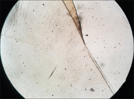
Figure 1: Edge-to-edge CEJ

 
thumb

Figure 2: Gap junction

 
thumb

Figure 3: Cementum overlapping enamel


Table 3: Percentage and frequency distribution of various types of cementoenamel junction in anterior and posterior teeth of males (sample size [n=60])

Types of CEJ Frequency, n (%)
Gap between enamel and cementum (Group 1) 21 (35)
Enamel overlapping cementum (Group 2) 0
Cementum overlapping enamel (Group 3) 4 (6.66)
Edge-to-edge contact (Group 4) 35 (58.33)

CEJ: Cementoenamel junction


In anterior and posterior teeth of females, the most common pattern observed was edge-to-edge type (55%), which was followed by gap between enamel and cementum (35%), cementum overlapping enamel (10%), and enamel overlapping cementum (0%) [Table 4].

Table 4: Percentage and frequency distribution of various types of cementoenamel junction in anterior and posterior teeth of females (sample size [n=60])

Types of CEJ Frequency, n (%)
Gap between enamel and cementum (Group 1) 21 (35)
Enamel overlapping cementum (Group 2) 0
Cementum overlapping enamel (Group 3) 6 (10)
Edge-to-edge contact (Group 4) 33 (55)

CEJ: Cementoenamel junction


DISCUSSION

The anatomy as well as arrangement of cementoenamel junction of permanent teeth possesses huge significance because of its association with various factors, for example, various pathological changes such as abrasion, cervical erosion, root surface caries, and resorption.

In the present study, a total number of 120 permanent teeth were selected, and ground section of all teeth was prepared. Later, the data were prepared and subjected to statistical analysis. Most of the data available regarding the cementoenamel junction demonstrate the presence of three morphological patterns (edge to edge, gap between the cementum and enamel, and cementum overlapping the enamel).[5-8]

The fourth type of morphology, i.e., enamel overlapping the cementum was defined by NeuvWald and Consolaro[9] in 2000 and Ceppi et al.[10] in 2006 in their research on permanent as well as deciduous teeth. A study done by Arambawatta et al.[2] found that edge-to-edge contact was more common in occurrence. In the present study, our observations were similar to that of the Arambawatta.

Bevenius et al.[8] conducted the study using eight recently extracted premolars and observed that edge-to-edge contact was the most common type of cementoenamel junction.

In theirstudy, Stosic[11] also observed that the most common pattern of the cementoenamel junction is edge-to-edge type. They also observed that in 33.3% of cases, cementum overlapping the enamel pattern wasseen, and 16.7% of cases showed enamel overlapping the cementum pattern. However, in our study, we have observed 6.66% and 0% cases showing cementum overlapping enamel and enamel overlapping cementum, respectively, in males, whereas in females, 10% and 0% of cases show cementum overlapping enamel and enamel overlapping cementum, respectively.

CONCLUSION

The present study shows various types of cementoenamel junction, which is an important anatomical and morphological structure in ground section of the teeth. In our study, the edge-to-edge pattern of the cementoenamel junction is the most common pattern observed, followed by gap, cementum over enamel, and enamel over cementum. Additional study with more number of samples will be helpful to get more accurate data and to analyze various pathological conditions in relation to the cementoenamel junction.

Financial support and sponsorship

Nil.

Conflicts of interest

There are no conflicts of interest.

REFERENCES

1. Francischone LA, Consolaro A. Morphology of the cementoenamel junction of primary teeth. J Dent Child (Chic) 2008;75:252-9. [Google Scholar] [PubMed]

2. Arambawatta K, Peiris R, Nanayakkara D. Morphology of the cemento-enamel junction in premolar teeth. J Oral Sci 2009;51:623-7. [CrossRef] [Google Scholar] [PubMed]

3. Hug HU, van 't Hof MA, Spanauf AJ, Renggli HH. Validity of clinical assessments related to the cemento-enamel junction. J Dent Res 1983;62:825-9. [CrossRef] [Google Scholar] [PubMed]

4. Avery JK, Chiego DJ Jr. Essentials of Oral Histology and Embryology. 3rd ed. USA:Elsevier;2006. [Google Scholar]

5. Hargreaves JA, Grossman ES, Matejka JM. Scanning electron microscopic study of prepared cavities involving enamel, dentin, and cementum. J Prosthet Dent 1989;61:191-7. [CrossRef] [Google Scholar] [PubMed]

6. Schroeder HE, Scherle WF. Cemento-enamel junction–revisited. J Periodontal Res 1988;23:53-9. [CrossRef] [Google Scholar] [PubMed]

7. Grossman ES, Hargreaves JA. Variable cementoenamel junction in one person. J Prosthet Dent 1991;65:93-7. [CrossRef] [Google Scholar] [PubMed]

8. Bevenius J, Lindskog S, Hultenby K. The amelocemental junction in young premolar teeth. A replica study by scanning electron microscopy. Acta Odontol Scand 1993;51:135-42. [CrossRef] [Google Scholar] [PubMed]

9. Neuvald L, Consolaro A. Cementoenamel junction:Microscopic analysis and external cervical resorption. J Endod 2000;26:503-8. [CrossRef] [Google Scholar] [PubMed]

10. Ceppi E, Dall'Oca S, Rimondini L, Pilloni A, Polimeni A. Cementoenamel junction of deciduous teeth:SEM-morphology. Eur J Paediatr Dent 2006;7:131-4. [Google Scholar] [PubMed]

11. Stosic N, Dacic S, Simonovic DD. Morphological variations of the cemento-enamel junction in permanent dentition. Acta Facultatis Medicae Naissensis 2015;32:209-14. CrossRef] [Google Scholar]