Department of Conservative Dentistry and Endodontics, Government Dental College and Hospital, Solan, Himachal Pradesh, India; 2Department of Prosthodontics, Bhojia Dental College, Solan, Himachal Pradesh, India; 3Department of Prosthodontics, Luxmi Bai Dental College, Patiala, Punjab, India; 4Department of Oral Surgery, Luxmi Bai Institute of Dental Sciences and Hospital, Patiala, Punjab, India; 5Tooth for Life Dental Clinic and Implant Center, Patiala, Punjab, India
Address for correspondence: Dr. Sumeeta Sandhu, House No. 79, Phase 3, Urban Estate, Patiala - 147 002, Punjab, India. E-mail: drsumeeta@gmail.com
DOI: 10.4103/2454-3160.202121
Saint Int Dent J 2016;2:7-10.
Copyright: © 2016 The Saint's International Dental Journal
Licence
This is an open access journal, and articles are distributed under the terms of the Creative Commons Attribution-NonCommercial-ShareAlike 4.0 License, which allows others to remix, tweak, and build upon the work non-commercially, as long as appropriate credit is given and the new creations are licensed under the identical terms.
Disclaimer:
This article was originally published by Wolters Kluwer Medknow Publications & Media Pvt Ltd and has now been officially transferred to Society of Dental Research & Education due to a change in publishing entity.
Healthy mouth needs a stable occlusal relationship to help in efficient mastication, maintaining vertical dimension of the face, stabilizing the arches, and protecting the soft tissues. Restorative dentistry loses its significance in the absence of good occlusion. Poorly occluding restored tooth surfaces not only provide a deleterious effect to the tooth and its associated periodontium but also to the dentition of the opposing arch. The damaging effects of premature contacts, cuspal interferences, faulty proximal contacts, etc., have an adverse influence on the overall health of stomatognathic system. In this article, the significance of establishing occlusal anatomy while restoring teeth to maintain healthy and harmonious occlusion has been described.
Key words: Axial loading, occlusal anatomy, occlusal forces
The subject of occlusion is such that it forms a medium to bring all the branches of dentistry together. This thought is made practically possible during clinical practice where all the elements of dentistry work together, aiming to perform a treatment that leaves the mouth in best biomechanical condition possible. Therefore, when we study occlusion, it should be viewed in a cohesive and comprehensive manner.[1] Occlusion is not just about teeth; it involves the entire stomatognathic system. It requires developing an understanding of the interrelationship between teeth, periodontal tissues, bones, joints, muscles, nervous system during the full range of mandibular movements.[2] During restorative procedures, it is important for every student of dentistry to have a knowledge of acceptable contact relationships of teeth and their influence on occlusal forces.
A healthy mouth is maintained in the best condition by relating cusps to condyles so that neither one adversely influences the other producing any type of havoc. Occlusal anatomy, however, complex it might appear to be, every complexity has a purpose of its own and a reason to be there. A restorative dentist must respect these complexities and the role they play in maintaining harmony in the overall system. While placing a restoration, a dentist must make an effort to create an occlusion that best suits with the oral environment. Cusps need to be properly placed in the interarch space and must be properly graded in heights and sizes to be in harmony with the movements of the mandible.[3] Optimal occlusion is that which is comfortably adapted by the patient. Therefore, a restoration is designed in a manner that it does not produce any disturbance in the normal condylar path. The magnitude and direction of load borne by the tooth is influenced by its occlusal anatomy.
In posterior teeth, the occlusal morphology is reproduced in a fashion that during mandibular closure there are even simultaneous contacts of maximum possible teeth achieving (a) maximal stability of the teeth (b) minimizing the amount of force placed on each tooth in function. The contact pattern of the teeth should be such that it directs the occlusal forces favorably for their adequate dissipation.
The tooth is housed in a bony socket with intervening periodontal ligament. The osseous tissue does not tolerate occlusal forces well; it shows resorption when under pressure. The periodontal ligament contains collagenous fibers that attach the tooth to the bony socket. Under the influence of occlusal forces, the fibers support the tooth, and tension gets created at the point of attachment of these fibers in the alveolar socket. This tension (i.e., pulling) actually stimulates osseous tissue formation. Therefore, a destructive force (i.e., pressure) is converted into a constructive force (i.e., tension) by the periodontal ligament, thereby acting as a natural shock absorber.
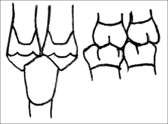
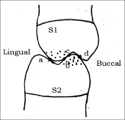
When the occlusal morphology is such that the cusp tip is contacted on a flat surface, i.e. bottom of a fossa or crest of the marginal ridge, the resultant occlusal force gets directed vertically along the long axis of the tooth. The fibers of periodontal ligament are arranged in a manner that such forces are well accepted and dissipated [Figure 1].
Figure 1: Cusp-fossa contact. Occlusal force (v) directed vertically along the long axes of teeth. Uniform tension (T) seen in periodontal ligament fibers
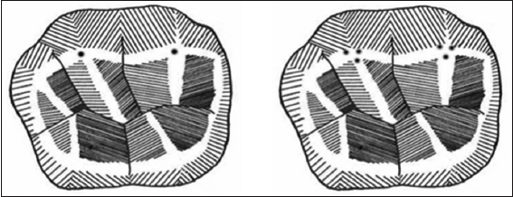
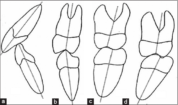
As opposed to this, when the occlusal morphology is such that the teeth contact each other on an incline, the resultant force is not directed along the long axis, instead a horizontal component is created which tends to cause tipping of teeth. The fibers of the periodontal ligament are not properly aligned to control them. On tipping, some fibers get compressed while others get pulled. Overall the forces are not effectively dissipated to the bone which can result in a pathological response of the bone in the form of resorption [Figure 2]. A neuromuscular reflex response may also be created in an attempt to avoid the incline contacts.[3,4] Improperly generated forces during incline contacts can have adverse influences such as tooth fracture, tooth jiggling, or mandibular deflection [Figure 3].[5]
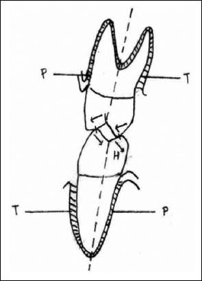
Figure 2: Cuspal incline contact. Horizontal component (H) of occlusal force created causing tipping of teeth. This tends to cause compression (P) and tension (T) in the periodontal ligament
Figure 3: Clinical Consequences of incline contacts (a) cuspal fracture, (b) tooth jiggling, (c) mandibular deflection
The process of directing occlusal forces through the long axis of the tooth is known as axial loading. It can be achieved by the following contact patterns:
Cusp - fossa/cusp – ridge contact
Tripod contacts.
Cusp - fossa occlusal relationship (tooth-to-tooth relationship) In cusp-fossa pattern of occlusion, the cusps and fossae of one tooth occlude with the cusps and fossae of only one opposing tooth [Figure 4].
Figure 4: Tooth-to-tooth relationship (cusp-fossa relationship)
In cusp-ridge pattern of occlusion, there is fitting of one stamp cusp into a fossa and fitting of another stamp cusp of the same tooth into the embrasure area of two opposing teeth (against two marginal ridges of adjacent teeth). It is the most stable relationship [Figure 5].[6]
Figure 5: Tooth-to-two teeth relationship (cusp-ridge relationship)
In complete closure in a normal young mouth, the contact point of the supporting cusp is neither at the very tip of the cusp and nor it is a single point. In fact, the supporting cusps are held in firm position by at least three contacts. These contacts occur on the inclines of the cusps. They are called tripod contacts, and the phenomenon is called tripodization.
They provide occlusal stability both buccolingually and mesiodistally. There is one buccal and one lingual contact for each cusp along with one mesial or distal contact. These contacts can be with opposing marginal, triangular, transverse, or oblique ridges. With advancing age, there occurs attrition and wear of dentition. The supporting cusps become more and more blunt and seat closer and closer to the bottoms of opposing fossae. Tripodization does not remain as prominent. Eventually, numerous flat cusps and surface contact are formed. These flat, smooth contacts do not allow any definite locking of the jaws which otherwise occurs [Figure 6].[7]
Figure 6: For cusp S1, contact points are a, b, c, For cusp S2 contact points are b, c, d
Thus, proper axial loading can be achieved when such contact patterns are produced as shown [Figure 7].
Figure 7: Contact patterns produced to achieve proper axial loading
To achieve proper axial loading the occlusal forces are directed along the long axes of the teeth, but the axes of maxillary and mandibular teeth have different angulations which vary with arrangement and location of teeth in the arch. These angulations also have to be borne in mind while developing restorations [Figure 8].
Figure 8: Different angulations of the teeth (a) incisors, (b) premolar, (c) first molar, (d) second molar
Anterior teeth are restored to their normal shape and contour. On mouth closing, no heavy contacts should be present between the upper and lower anterior teeth. These tend to displace the teeth anteriorly or cause heavy vibration known as fremitus. 8]
To summarize, the restorations should be such that during buccal, lingual, anteroposterior or any other mandibular border movements, the upper and lower teeth should move over their opponents without collisions or contacts and during intercuspation they should thrive to achieve maximal occlusal stability.
Mouth like any other organ in the body is a self-sufficient unit. There exists a protective relationship between the anterior and posterior teeth which should be respected and not violated while performing restorative procedures. Placement of restorations should not lead to the generation of any abnormal forces or creation of lateral stresses. In fact, they should be so well integrated in the system that a feeling of artificial replacement does not occur.
Nil.
There are no conflicts of interest.
1. Mohl ND, Zarb GA, Carlsson GE, Rugh JD. A Textbook of Occlusion. 1st ed. Chicago, Illinois:Quintessence Publishing Co. Inc.;1988. [Google Scholar]
2. Davies S, GrayRM. What is occlusion?Br Dent J 2001;191:235-8, 241-5. [CrossRef] [Google Scholar] [PubMed]
3. Stuart CE. Why dental restorations should have cusps. J Prosthet Dent 1960;10:553-5. [CrossRef] [Google Scholar]
4. Okeson JP. Management of Temporomandibular Disorders and Occlusion. 6th ed. CV Mosby Company, St. Louis (MO):2008. [Google Scholar]
5. Davies SJ, Gray RM, Smith PW. Good occlusal practice in simple restorative dentistry. Br Dent J 2001;191:365-8, 371-4, 377-81. [CrossRef] [Google Scholar] [PubMed]
6. Roberson TM, Heyman HO, Swift EJ. Strudevant's Art and Science of Operative Dentistry. 5th ed. Pheladelphia:Mosby Inc.;2006. [Google Scholar]
7. Ash MM, Ramfjord S. Occlusion. 4th ed. Philadelphia-London-Tronto:WB Saunders Company;1995. [Google Scholar]
8. Howat AP, Capp NJ, Barrett NV. A Colour Atlas of Occlusion and Malocclusion. 1st ed. London, England:Wolfe Publishing Ltd.;1991. [Google Scholar]