Keratocystoma of minor salivary gland of the palate

Gourav Ahuja, Kamaldeep Kaur Aulakh1, Eish Sethi2, Varun Nagpal3, Harkanwal Kaur1

Department of Oral and Maxillofacial Surgery, B.R.S. Dental College and Hospital, Panchkula, Haryana, India; 2Department of Oral and Maxillofacial Surgery, Dasmesh Institute of Research and Dental Sciences, Faridkot, Punjab, India; 3Department of Oral and Maxillofacial Surgery, Desh Bhagat Dental College and Hospital, Sri Muktsar Sahib, Punjab, India; 4Department of Oral and Maxillofacial Surgery, Jan Nayak Ch. Devi Lal Dental College, Sirsa, Haryana, India


Address for correspondence: Dr. Gourav Ahuja, Department of Oral and Maxillofacial Surgery, B.R.S. Dental College and Hospital, Panchkula, Haryana, India. E-mail: ahujaos34@gmail. com


DOI: 10.4103/2454-3160.202125

Saint Int Dent J 2016;2:21-23.

Copyright: © 2016 The Saint's International Dental Journal

Licence

This is an open access journal, and articles are distributed under the terms of the Creative Commons Attribution-NonCommercial-ShareAlike 4.0 License, which allows others to remix, tweak, and build upon the work non-commercially, as long as appropriate credit is given and the new creations are licensed under the identical terms.

Disclaimer:

This article was originally published by Wolters Kluwer Medknow Publications & Media Pvt Ltd and has now been officially transferred to Society of Dental Research & Education due to a change in publishing entity.


Abstract:

The authors analyzed the clinical, histopathological, and immunohistochemical findings of a slowly growing palatal gland tumor in a 34-year-old female. The cut surface of the excised mass showed multilocular cystic spaces with grayish brown solid areas. Histological findings demonstrated a benign tumor with multicystic spaces and focal solid epithelial cells lined by benign squamous epithelial cells. The overlying epithelium was parakeratinized and without a granular layer. The cystic spaces were filled with keratin material. On immunohistochemistry examination, the tumor cells were found positive for HMWCK, focally for CK7, CK19 and p63 and Mib-1 labeling was approximately 1%. There was no evidence of recurrence after 8 months of resection. The data from this case and previously published studies suggest that keratocystoma of both major and minor salivary glands is a benign neoplasm and that surgical resection is apparently a sufficient treatment modality.

Key words: Choristoma, keratocystoma, rare benign tumor


Of all the tumors of salivary glands, keratocystoma is a relatively new, rare, benign entity.[1] The pathognomonic features of keratocystoma include multicystic spaces lined with stratified squamous cells and the presence of focal solid epithelial nests.[2]

We reviewed internet-based databases such as PubMed and MEDLINE using the keywords choristoma, rare benign salivary gland tumor, and keratocystoma for previously published reports/ studies of keratocystoma. To best of our knowledge, only eight cases occurring exclusively in the parotid gland have been published till date.[3-7] These cases involved children and adults (aged between 8 and 49 years). In all of these, the treatment modalities varied from superficial to subtotal parotidectomy along with facial nerve exclusion. There has been no evidence of recurrence in the postoperative follow-ups in any of these cases.

The authors claim to have found the first case of keratocystoma presenting at an unusual site, i.e. the palate. Here, they present the clinical, histopathological, and immunohistochemistry (IHC) features of this case; discussion of earlier reported radiological results and a review; and clinicopathological findings [Table 1].

Table 1: Review of clinicopathological findings of keratocystoma

Authors Year Age/sex Site/side Associated symptoms Treatment modality Follow-up and rate of recurrence
Seifert et al .[4] 1999 8/female Parotid gland/right None No detail No detail
Nagao et al.[1] 2002 18/male Parotid gland/right None Subtotal parotidectomy 3 years/none
Nagao et al.[1] 2002 38/male Parotid gland/left None Subtotal parotidectomy 2 years/none
Zhang et al[5] 2010 37/male Parotid gland/left None Subtotal parotidectomy 18 months/none
Huang[6] 2012 29/female Parotid gland/right None Subtotal parotidectomy 4 years/none
Huang et al.[6] 2012 49/female Parotid gland/right None Subtotal parotidectomy 3 years/none
Hirata[2] 2014 34/male Parotid gland/left Tender on palpation Subtotal parotidectomy 17 months/none
Wang et al.[3] 2015 34/male Parotid gland/left None Superficial parotidectomy 7 years/none
Present case 2016 34/female Left posterior soft palate None Surgical excision with safe margin of 0.5 cm 8 months/none
 

CASE REPORT

A 34-year-old female presented with a painless, submucosal, movable mass in the soft palate which was present since 3 months. The mass was located in the left posterior palatal region, measuring about 2 cm × 1.5 cm in size with a distinguishable boundary [Figure 1a]. The mass was oval in shape, firm in consistency, and nonpulsatile in nature with overlying mucosa normal in color and texture. There was no complaint of paraesthesia. The clinical features simulated a benign lesion, and so an excisional biopsy was planned. A signed informed consent was taken from the patient for the surgical intervention which was performed keeping a safe margin of 0.5 cm [Figure 1b].

thumb

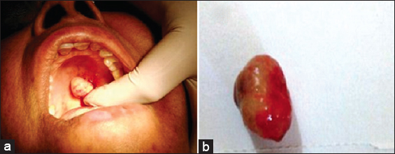
Figure 1: (a) Clinical presentation of keratocystoma of the palate. (b) Resected specimen of keratocystoma


Macroscopically, the cut surface of the excised mass demonstrated multilocular cystic spaces showing grayish brown solid areas. Histologically, the section showed a benign tumor with multicystic spaces and focal solid epithelial cells lined by benign squamous epithelial cells but without lobar architecture [Figure 2a]. The overlying epithelium was parakeratinized and without a granular layer. Cells had abundant eosinophilic cytoplasm with bland, uniform nuclei [Figure 2b]. The cystic spaces were filled with keratin material. These findings helped us to reach a provisional diagnosis of keratocystoma. However, keeping in mind the rarity and uncertain behavior of this lesion and to confirm the diagnosis, we went for advanced immunohistochemical study with a panel of markers (high molecular weight cytokeratin, cytokeratin, p63, and MiB-1). On IHC examination, the tumor cells were found positive for HMWCK, focally for CK7, CK19, and p63 and Mib-1 labeling was approximately 1% [Figure 3a-c]. Furthermore, focal foreign body giant cell reaction to the keratin material was visible. The histological and IHC findings were consistent with those of keratocystoma which suggested of a diagnosis of keratocystoma of the palate.

thumb

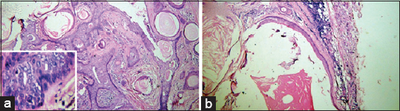
Figure 2: (a) Photomicrograph showing multicystic spaces and numerous keratin pearl like areas (H and E stain, ×10); Inset (×40) showing cells with eosinophilic cytoplasm and bland uniform nuclei. (b) Photomicrograph showing parakeratinized cystic lining and epithelium (H and E stain, ×10; scanner view)

 
thumb

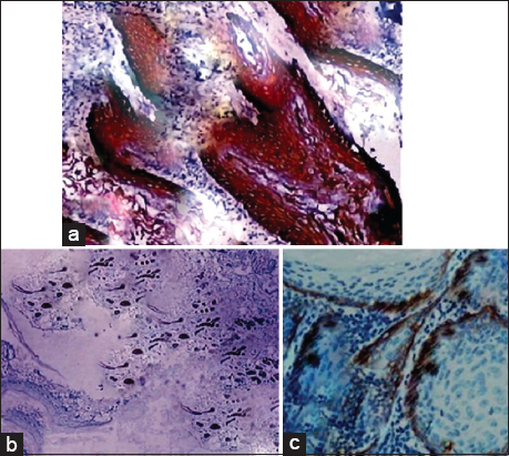
Figure 3: (a-c) Immunohistochemical findings of specimen. (a) Tumor cells are positive for HMWCK. (b) Focally for CK7 and (c) p63


DISCUSSION

Seifert et al.[4] published a case report of an unusual benign tumor of the parotid gland which had a histopathological finding of the presence of multicystic spaces lined by stratified squamous cells containing keratotic lamellae with focal solid epithelial nests. Moreover, it was accompanied by foci of calcification and foreign body reaction. The authors placed this tumor in the category of “choristoma” pertaining to its resemblance with “trichoadenoma” of skin.[2] Thereafter, Nagao et al.[1] reported two cases of painless swellings of the parotid gland. They observed certain microscopic findings such as(1) bland appearing nuclear morphology of the squamous cells, (2) simultaneous metaplasia of ducts and acini, (3) conspicuous inflammatory constituents and granulation tissue, (4) development of larger cysts, and (5) lack of granular layer in the squamous lining. The authors believed that this lesion was of a peculiar variety of salivary gland tumors and hence proposed a new designation, “keratocystoma,” to present its specific histopathological features. In addition, they stated that these features might be helpful in distinguishing it from necrotizing metaplasia.

Zhang et al.[5] described a painless circumscribed nodule of the parotid gland as keratocystoma based on the histopathological findings similar to those of earlier reports. Moreover, from IHC examination, the tumor cells were diffusely positive for CKAE1/AE3 and 5/6 and focally for CK 7, stained strongly for p63 but weakly for p53. In the year 2012, X-F Huang et al.[6] reported two cases of keratocystoma in two women aged 29- and 49-year-old suggesting a broader age range for the occurrence of this lesion. Both cases immunoreactivity stained positively for AE1/AE3 and CK5/6 but negatively for CK8/18, S-100, and calponin. The authors also added a new peculiar histopathological finding, i.e. the sporadic presence of basophilic keratohyalin granules in the orthokeratinized squamous epithelium of this tumor.

Radiological imaging of keratocystoma recorded with ultrasound, magnetic resonance imaging (MRI), and salivary scintigraphy was reported by Hirata et al.[7] The ultrasound images produced after injection of gadolinium contrast medium (0.2 mmol/kg) proposed this lesion as pleomorphic adenoma pertaining to the fact that it had a thin, homogenous wall that was enhanced by the contrast medium. However, the MRI findings differed from the characteristic findings of pleomorphic adenoma-like solid component with intensive enhancement. To add to it, the authors observed a ring-like enhancement of the lesion wall which indicated a viable cell component. Salivary scintigraphy was performed using 99mTcO4, but the tracer element did not accumulate in the tumor indicating that the mass did not contain any saliva. And thereby, the authors suggested that salivary scintigraphy is not useful for diagnosing keratocystoma as there are many other salivary gland tumors which also show the absence of uptake of the tracer element.

Recently, a case report of a keratocystoma of the parotid gland was published by Wang et al.[3] They observed a low index of Ki-67 restricted to basal layer of cysts and nests from immunohistochemical examination, a benign cytological morphology from histopathological reports and a relatively slow, painless clinical course of the lesion which confirmed the benign nature of keratocystoma. They also stated that the absence of mucus cells and extensive keratinization helped in ruling out mucoepidermoid carcinoma. In addition, the lack of a granular layer in the multicystic spaces in a keratocystoma had a major role in distinguishing it from a benign tumor like an epidermoid cyst or a trichoadenoma.

CONCLUSION

Keratocystoma has not been listed as an independent pathological entity in the revised WHO classification of salivary gland tumors (3rd edition) but as a tumor in the differential diagnosis of squamous cell carcinoma of salivary glands.[8,9] The above case report discusses a rare case of keratocystoma of minor salivary glands of the palate. Data from earlier publications revealed that this benign tumor occurred in major salivary glands, i.e. parotid gland and hence the designation of keratocystoma of the parotid gland was introduced as a newly recognized diagnosis.[10] Herein, we highlight the need for studies regarding site predilection of this tumor are required to categorize keratocystoma as a benign tumor of both minor and major salivary glands.

Financial support and sponsorship

Nil.

Conflicts of interest

There are no conflicts of interest.

Acknowledgment

The authors would like to acknowledge Altus Laboratories, Chandigarh, and Tata Memorial Hospital, Mumbai, Maharashtra, India.

REFERENCES

1. Nagao T, Serizawa H, Iwaya K, Shimizu T, Sugano I, Ishida Y, et al. Keratocystoma of the parotid gland:A report of two cases of an unusual pathologic entity. Mod Pathol 2002;15:1005-10. [CrossRef] [Google Scholar] [PubMed]

2. Kumar V, Fausto N, Abbas A. Neoplasia. Robbins Pathologic Basis of Disease. 7th ed. Philadelphia:WB Saunders;2004. 6. [Google Scholar]

3. WangW, LiY, TangY. Keratocystoma of the parotid gland:A case report and review of previous publications. Br J Oral Maxillofac Surg 2015;53:655-7. [CrossRef] [Google Scholar] [PubMed]

4. Seifert G, Donath K, Jautzke G. Unusual choristoma of the parotid gland in a girl. A possible trichoadenoma. Virchows Arch 1999;434:355-9. [CrossRef] [Google Scholar] [PubMed]

5. Zhang MF, Lin SX, Yi C, Fu J, Wu QL, Yun JP. Keratocystoma of the parotid gland:Case report and immunohistochemical investigation. J Clin Pathol 2010;63:758-60. [CrossRef] [Google Scholar] [PubMed Central] [PubMed]

6. Huang XF, LiW, Yang XD, JiangWH, Chen XH, Hu QG. Keratocystoma of the parotid gland:A clinicopathological study and literature review. Int J Oral Maxillofac Surg 2012;41:256-60. [CrossRef] [Google Scholar] [PubMed]

7. Hirata K, Fujima N, Mizumachi T, Bandarchi B, Roesler JM. Radiological images of keratocystoma:A rare tumor of the parotid gland. Acta Radiol Short Rep 2014;3:2047981614549497. [CrossRef] [Google Scholar] [PubMed Central] [PubMed]

8. Lewis JE, Olsen KD. Tumours of the salivary glands. In:Barnes L, Eveson JW, Reichart P, Sidransky D, editors. Pathology and Genetics of Head and Neck Tumours. World Health Organization Classification of Tumours. 3rd ed. Lyon:International Agency for Research on Cancer (IARC) Press;2005. 246. [Google Scholar]

9. Barnes L, Eveson JW, Reichart P, Sidransky D. Pathology and Genetics of Head and Neck Tumours. World Health Organization Classification of Tumours. Lyon:IARC Press;2005. Google Scholar] [PubMed Central] [PubMed]

10. Cheuk W, Chan JK. Advances in salivary gland pathology. Histopathology 2007;51:1-20. [Google Scholar] [CrossRef] [PubMed]