Retention of various overdenture posts: An in vitro study

Sanam Kumar, Babita Karda1, Kavipal Singh2, Nimish Sethi2

Department of Dental, Civil Hospital, Hoshiarpur, India, 1Department of Pedodontic and Preventive Dentistry, Desh Bhagat Dental College and Hospital, Muktsar, India, 2Department of Prosthodontics and Crown and Bridges, Sri Guru Ram Das Dental College and Hospital, Amritsar, Punjab, India


Address for correspondence: Dr. Babita Karda, Street No. 1B, New Gobind Nagar, B/H – Sonalika Threshers, Jalandhar Road Hoshiarpur - 146 001, Punjab, India. E-mail: dr_babita08@ yahoo.co.in


DOI: 10.4103/2454-3160.177945

Saint Int Dent J 2015;1:112-6.

Copyright: © 2015 The Saint's International Dental Journal

Licence

This is an open access journal, and articles are distributed under the terms of the Creative Commons Attribution-NonCommercial-ShareAlike 4.0 License, which allows others to remix, tweak, and build upon the work non-commercially, as long as appropriate credit is given and the new creations are licensed under the identical terms.

Disclaimer:

This article was originally published by Wolters Kluwer Medknow Publications & Media Pvt Ltd and has now been officially transferred to Society of Dental Research & Education due to a change in publishing entity.


Abstract:

Context Tooth retained overdenture helps to reduce the impact of some of the complete denture wearing consequences: Residual ridge resorption, loss of occlusal stability, undermined esthetic appearance, and compromised masticatory function.

Aims This in vitro study compared the retention of three prefabricated overdenture posts cemented with self-adhesive resin cement.

Subjects and Methods Thirty freshly extracted noncarious, healthy mandibular canines were sectioned 1 mm above cementoenamel junction and endodontically treated.

The teeth were divided in three groups and prepared for overdenture posts Group 1: Access post overdenture (EDS, USA), Group 2: Flexi overdenture post (EDS, USA), and Group 3: Ceka Preci-Clix overdenture (Ceka Preci-line, Germany). Posts were cemented with self-adhesive resin cement (Relyx U 100, Germany). Each tooth was positioned identically in the universal testing machine (Instron), and the amount of force necessary to remove a post from the tooth was recorded.

Statistical Analysis Used: The results obtained were statistically analyzed using one-way analysis of variance and the comparisons of groups were performed using post hoc Tukey honestly significant difference test.

Results Ceka Preci-Clix overdenture post has maximum mean retention value of 171.57N and access post overdenture has minimum retention value of 98.75N.

Conclusions Ceka Preci-Clix overdenture post was more retentive (171.57) due to its parallel sided design than the other posts used in this study. Flexi overdenture post, due to its split shank threaded design, was the 2nd most retentive post (128.88) after Ceka Preci-Clix overdenture post and access post overdenture were least retentive post (98.75) due to its thick-walled hollow design.

Keywords: Access post, Ceka Preci-Clix post, Flexi post, overdenture post, self-adhesive resin cement, universal testing machine


In the rehabilitation of edentulous patients, the clinician faces the problem of greatly resorbed ridges, excess salivary flow, reduced muscle tone, and other factors, which lead to decreased retention and support of the prosthesis.[1] One therapeutic approach directed at improving oral function in elderly edentulous patients is the use of overdentures.[2]

Survival of the overdenture post placed in natural teeth depends upon the ability of the posts to resist the functional force of mastication as well as stress created during insertion and removal of the denture that can lead to loosen or displacement of the post from the canal.[3]

AIMS AND OBJECTIVE

The aim of this study was to compare the retention of three prefabricated overdenture posts: Access post overdenture [EDS, USA Figure 1], Flexi overdenture post [EDS, USA Figure 2], Ceka Preci-Clix overdenture post [Ceka Attachments Preci-Line, Germany, Figure 3] cemented with self-adhesive resin cement [Relyx U 100, 3M ESPE, Germany, Figure 4].

thumb

Figure 1. Access post overdenture

 
thumb

Figure 2. Flexi overdenture post

 
thumb

Figure 3. Ceka Preci-Clix overdenture post

 
thumb

Figure 4. Relyx U 100


SUBJECTS AND METHODS

The present study was carried out on thirty freshly extracted noncarious, healthy mandibular canines of comparable root length. Samples collected were stored in 0.5% sodium hypochlorite solution. All the samples were sectioned 1 mm above the cementoenamel junction with the help of diamond disk attached to straight handpiece [Figure 5] and then the samples were endodontically treated. Radiograph of the tooth was taken to determine the length of the root. Access to root canal was made with round bur attached to air-rotor handpiece (NSK). Necrotic pulp tissue was removed with the help of barbed broach (Dentsply). Debridement of the canal was done with K files (Dentsply) and Reamers (Dentsply), and sodium hypochlorite was used to irrigate the canal to remove the rest of infected tooth structure. The root canal was dried with the help of paper points (Dentsply). Obturation of the canal was done with sectional condensation method. A gutta-percha cone (Dentsply, Maillefer) of approximately the size of prepared canal was cut into sections each of 3 or 4 mm long. Root canal plugger (Dentsply, Maillefer) was selected that can be inserted in the canal to within 3–4 mm of apex. The canal was coated with zinc oxide eugenol cement (GC, Japan). Root canal plugger was heated in the salt hot sterilizer for 10 s and cut apical section of selected gutta-percha cone was mounted on heated plugger and carried into root canal to measure depth and pressed vertically. The plugger was disengaged very carefully to prevent dislodging of the inserted section of gutta-percha. After obturation, minimum of 9 mm of root canal space was available. Then the radiograph was taken to check the position and fit of condensed section [Figure 6]. All the samples were mounted in polyvinyl chloride ring using polymethyl methacrylate with cementoenamel junction 1mm above the top of the ring in its most vertical alignment with the help of prosthodontic surveyor. The specimens were randomly divided into three groups containing 10 specimens each. In Group 1, access post overdenture was used, post space was prepared with access post overdenure reamer, and countersink drill provided with access post overdenture kit was used to prepare post space for the second tier and flange portion of the overdenture post. In Group 2, Flexi overdenture post was used, post space was prepared with Flexi overdenture reamer and countersink drill provided with Flexi overdenture kit was used to prepare post space for the second tier and flange portion of the overdenture post. In Group 3, Ceka Preci-Clix overdenture post was used. First root canal was prepared with Ceka Preci-Clix penetration bur after that diamond bur provided with Ceka Preci-Clix overdenture post kit was used to prepare the base of Preci-Clix overdenture post. Finally, Ceka Preci-Clix overdenture reamer was used to calibrate the diameter for the post [Figure 7]. All the posts were luted with self-adhesive resin cement Relyx U 100. Posts were held under finger pressure for 5 min till the initial set of cement. Retention of overdenture post of each sample was tested with the universal testing machine (model LR – 100, 1998, INSTRON, UK), [Figure 8] at a crosshead speed of 1 mm/min. Acrylic resin block was mounted firmly to lower jaw of the universal testing machine (model LR – 100, 1998,

thumb

Figure 5. Sectioning of tooth 1mm above cementoenamel junction

 
thumb

Figure 6. Radiograph to check position and fit of condensed section

 
thumb

Figure 7. Prepared post space

 
thumb

Figure 8. Universal testing machine


INSTRON, UK) and the overdenture post was connected to the upper jaw. The samples were subjected to an axial dislodgment, and the force in Newton was recorded when separation was observed in the universal testing machine. The result thus obtained was statistically analyzed using one-way analysis of variance (ANOVA) and the comparisons of groups were performed using post hoc Tukey honestly significant difference (HSD) test.

RESULTS

Table 1 shows mean tensile strength (Newton) for all the three groups. The mean tensile strength was highest for Group 3 - Ceka Preci-Clix overdenture post (171.572 ± 11.51N) followed by Group 2 - Flexi overdenture post (128.883 ± 18.36N). The mean tensile strength was lowest for Group 1 access post overdenture (98.758 ± 10.05N).

Table 1. Descriptive average tensile strength (Newton) of all the groups

Group n Range Mean±SD 95% CI
Group 1 - Access post overdenture 10 78.84-111.79 98.758±10.049 91.569-105.947
Group 2 - Flexi overdenture post 10 107.96-158.08 128.883±18.359 115.750-142.016
Group 3 - Ceka Preci-Clix overdenture post 10 157.58-190.24 171.572±11.508 163.339-179.804

SD = Standard deviation, CI = Confidence interval


The mean tensile strength in three groups was compared using one-way ANOVA with post hoc Tukey HSD.

Table 2 shows one-way ANOVA analysis done to compare inter and intragroup mean tensile strength. The mean tensile strength is statistically significant between the group (P < 0.001) with F value 70.396, whereas the values of mean tensile strength are statistically nonsignificant within the group.

Table 2. One-way analysis of variance for comparison within group and between groups

Sum of squares df Mean square F P Significance
Between groups (intergroup) 26,772.483 2 13,386.242 70.396 Highly significant Highly significant
Within groups (intragroup) 5134.238 27 190.157
Total : 31,906.721 29
 

Table 3 shows multiple comparisons for all three groups. When each group was compared with other using post hoc Tukey HSD, it showed that the mean difference between the tensile strength of Group 1 from Group 2 was 30.125 with P < 0.001, i.e., highly significant. Thus, the mean tensile strength of Group 1 differs significantly from Group 2. The mean difference between the tensile strength of Group 1 from Group 3 is 72.814 with P < 0.001, i.e., highly significant. Thus, the tensile strength of Group 1 differs highly significantly from Group 3. The mean difference between the tensile strength of Group 2 from Group 3 is 49.689 with P < 0.001, i.e., highly significant. Thus, the tensile strength of Group 2 differs highly significantly from Group 3.

Table 3. Comparison between the groups using post hoc Tukey honestly significant difference

Comparison Mean difference P Significance
Group 1 versus 2 30.125 <0.001 Highly significant
Group 1 versus 3 72.814 <0.001 Highly significant
Group 2 versus 3 42.689 <0.001 Highly significant
 

DISCUSSION

Resorbed residual ridges, excess salivary flow, reduced muscle tone, and other factors, pose a great challenge in the complete denture construction.[1] Variations in mandibular osseous and mucosal anatomy and structure, opposing maxillary dentition and/or restorations, alterations of temporomandibular and occlusal relationships, loss of vertical dimension of occlusion, material dynamics, and patient expectations present the dentist with complex combination of variables, resulting in the necessity for accurate diagnosis and treatment planning. Elderly patients often have progressively decreasing neurophysiologic adaptive capacity to wear complete denture with increasing age. Deteriorating muscle strength and coordination in elderly patients may lead to problems in fabricating complete denture as well as difficulty in achieving and maintaining acceptable denture stability and retention. Oral therapeutic approach directed at improving oral function in elderly edentulous patients is the use of overdenture.[2] Overdenture is defined as removable partial denture or complete denture that covers and rests on one or more remaining natural teeth, the roots of natural teeth and/or dental implants.

Tooth retained overdenture helps to reduce the impact of some of complete denture wearing consequences: Residual ridge resorption, loss of occlusal stability, undermined esthetic appearance, and compromised masticatory function.[4]

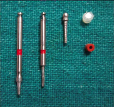
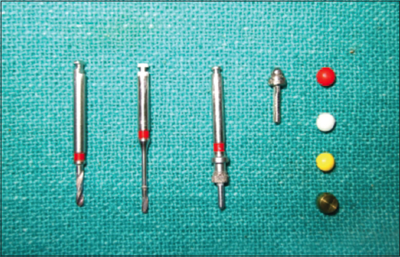
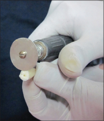
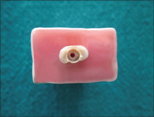
When an overdenture attachment is used, the available interocclusal distance of a standard denture cannot be compromised and so a struggle ensues to place all of the overdenture attachments within its proper dimension. Overdenture attachments are bulky in size, which may lead to weaker structure, which is difficult to clean and maintain. To overcome interocclusal space problem, overdenture posts are used to retain complete denture. These posts derive their retention within roots so can be used in inadequate interocclusal space. Other advantages of overdenture posts are: The leverage on the abutment tooth is negligible because the point of attachment is actually below alveolar bone level, and overdenture post system is simple to use, can be placed quickly at chair side, and can be done without any casting.[5] Among the various overdenture post designs, ball and socket design is the most acceptable. The ball and socket combination usually consists of one part of metal (usually the post) and other part made of nylon or plastic (cap or keeper is usually placed in denture). Wear caused by daily activities such as placement and removal of denture and normal oral environment may result in failure of the nylon or plastic. The wear of cap reduces the retention of the cap over its lifetime. However, inner plastic cap inserted into a metal keeper design allows the dentist to replace the worn out inner cap in seconds with the help of special wrench, which can be used to unscrew and remove the old (worn) cap and replace it with a new cap.[4] There are many variables to consider when choosing a prefabricated post overdenture attachment system such as the length of the retained root or roots (crown/root ratio), its configuration, and the quality and quantity of alveolar bone.[5]

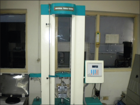
Results of this study showed that Ceka Preci-Clix overdenture post (Group 3) has maximum mean retention value of 171.57N [Table 1] as compared to mean retention value 128.88N [Table 1] of Flexi overdenture post (Group 2). Access post overdenture (Group 1) has minimum retention value of 98.75N [Table 1].

Maximum retention found in Ceka Preci-Clix overdenture post may be due its parallel-sided and threaded design. Flexi overdenture post has unique threaded split shank design that provides more retention without contributing to the production of tensile stress to root.[6] Retention of access post overdenture system is less due to its thick-walled hollow tube design, which offers the ability to remove a post in case of failed root canal, without surgically widening the canal.[7]

Kurer et al. studies found that parallel sided dowels were more retentive than tapered ones. Roughness or serrations on the surface increased axial retention. They found that increased length improved retention for all dowels tested.[8]

Deutsch et al. concluded that the most retentive posts in decreasing order are parallel threaded and then radix, parallel serrated, parallel smooth, and smooth wedge-shaped posts. Increased post length gave increased retention. Cement used and diameter of the post had little effect on retention.[9]

Flexi post by Musikant and Deutsch produced limited stress even under maximum torquing.[10]

Deutsch et al. showed that Flexi post had a greater retentive value than the other posts. It was concluded that the retention of the Flexi post became greater as the size of the post increased.[11]

A study conducted by Epstein et al. was in favor of the present study in which they compared retentive properties of six prefabricated overdenture post attachment system (access post overdenture, era white and era gray, Flexi overdenture post, o-so and zaag) and concluded that retention of access post overdenture was significantly lower than Flexi overdenture post. The reason for this may be that Flexi post has threaded split shank design, and threads are extremely sharp and cut deep into dentin rather than displace it.[4]

Leung and Preiskel confirmed concluded that mean retentive force for Flexi post overdenture is maximum as compared to other systems.[12]

Qualtrough et al. concluded that parallel sided light posts were significantly more retentive than parapost, fiber white posts, light post, and snow post. Of the other posts.[13]

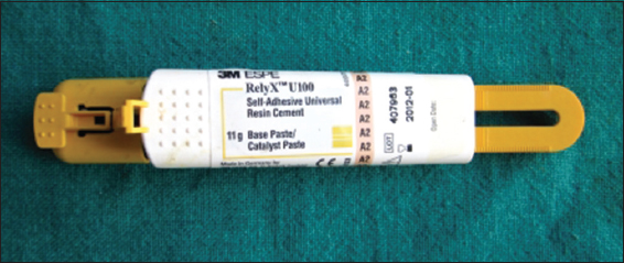
Testing here was directed at limited, specific, and expected mechanical conditions and this in vitro protocol undoubtedly falls short of clinical reality. Direct tensile test used in this study does not closely mimic what actually takes place in the oral environment under conditions of mastication, where overdenture posts would be subjected to repeated compressive loading that may lead to fatigue with subsequent failure. Nevertheless, it provided a vivid indication and ranking of retention value of three prefabricated overdenture posts cemented with self-adhesive resin cement.

CONCLUSION

This in vitro study was performed on 30 extracted mandibular canines. The retention of three prefabricated overdenture posts: Access post overdenture (EDS, USA), Flexi overdenture post (EDS, USA), and Ceka Preci-Clix overdenture (Ceka Preci-line, Germany), cemented with self-adhesive resin cement (Relyx U 100, Germany) was quantitatively determined. The teeth were divided into three groups containing ten samples each and prepared for overdenture posts. Each tooth was positioned identically in the universal testing machine (Instron), and the amount of force necessary to remove a post from the tooth was recorded.

Within limitations of study, it was concluded that Ceka Preci-Clix overdenture post was more retentive (171.57) due to its parallel sided design than the other posts used in this study. Flexi overdenture post due to its spilt shank threaded design was the 2nd most retentive post (128.88) after Ceka Preci-Clix overdenture post and access post overdenture was a least retentive post (98.75) due to its thick-walled hollow design.

Financial support and sponsorship

Nil.

Conflicts of interest

There are no conflicts of interest.

REFERENCES

1. Svetlize CA, Bodereau EF Jr. Comparative study of retentive anchor systems for overdentures. Quintessence Int 2004;35:443-8. [CrossRef] [Google Scholar] [PubMed]

2. Chung KH, Chung CY, Cagna DR, Cronin RJ Jr. Retention characteristics of attachment systems for implant overdentures. J Prosthodont 2004;13:221-6. [CrossRef] [Google Scholar]

3. Elsayed ME, El-Mowafy O, Fenton A. Retention of overdenture posts cemented with self-adhesive resin cements. Int J Prosthodont 2009;22:287-9. [CrossRef] [Google Scholar] [PubMed]

4. Rutkunas V, Mizutani H. Retentive and stabilizing properties of stud and magnetic attachments retaining mandibular overdenture –An in vitro study. Stomatologija Balt Dent Maxillofac J 2004;6:85-90. [CrossRef] [Google Scholar]

5. Epstein DD, Epstein PL, Cohen BI, Pagnillo MK. Comparison of the retentive properties of six prefabricated post overdenture attachment systems. J Prosthet Dent 1999;82:579-84. [CrossRef] [Google Scholar] [PubMed]

6. Sheldon W. Essentials of Complete Denture Prosthodontics. 2nd ed. India:A.I.T.B.S. Publishers;1979. 384. [CrossRef] [Google Scholar]

7. Cohen BI, Pagnillo M, Condos S, Deutsch AS. Comparative study of two precision overdenture attachment designs. J Prosthet Dent 1996;76:145-52. [CrossRef] [Google Scholar]

8. Kurer HG, Combe EC, Grant AA. Factors influencing the retention of dowels. J Prosthet Dent 1977;38:515-25. [CrossRef] [Google Scholar] [PubMed]

9. Deutsch AS, Musikant BL, Cavallari J, Lepley JB. Prefabricated dowels:A literature review. J Prosthet Dent 1983;49:498-503. [CrossRef] [Google Scholar] [PubMed]

10. Musikant BL, Deutsch AS. A new prefabricated post and core system. J Prosthet Dent 1984;52:631-4. [CrossRef] [Google Scholar] [PubMed]

11. Deutsch AS, Musikant BL, Cavallari J, Bernardi S. Retentive properties of a new post and core system. J Prosthet Dent 1985;53:12-4. [CrossRef] [Google Scholar] [PubMed]

12. Leung T, Preiskel HW. Retention profiles of stud-type precision attachments. Int J Prosthodont 1991;4:175-9. [CrossRef] [Google Scholar] [PubMed]

13. Qualtrough AJ, Chandler NP, Purton DG. A comparison of the retention of tooth-colored posts. Quintessence Int 2003;34:199-201. [CrossRef] [Google Scholar] [PubMed]